A Case of Valganciclovir Treatment for Cytomegalovirus Retinitis
- Affiliations
-
- 1Department of Ophthalmology, Inha University School of Medicine, Incheon, Koera.
- 2Ophthalmologic Oncology Clinic, National Cancer Center, Gyeonggi, Korea. dreye@freechal.com
- KMID: 2211331
- DOI: http://doi.org/10.3341/jkos.2008.49.3.531
Abstract
-
PURPOSE: To report a case of a patient with cytomegalovirus (CMV) retinitis who was treated with oral valganciclovir.
CASE SUMMARY
A 34-year-old man who had undergone anti-cancer chemotherapy for Non-Hodgkin lymphoma was referred to the ophthalmologic oncology clinic because of decreased vision in both eyes. Fundus examination showed white, opaque, and granular retinal lesions in both eyes, and a serologic test showed a positive response to CMV antibody IgG and a negative response to CMV antibody IgM. The patient received induction therapy with intravenous ganciclovir and maintenance therapy with oral valganciclovir 900 mg once daily. CMV retinitis reactivated 4 weeks after maintenance therapy was discontinued. At that point, the patient received induction therapy with oral valganciclovir 900 mg twice daily for 3 weeks and maintenance therapy with 900 mg once daily for 5 weeks. The retinal lesion disappeared and did not recur after oral administration of valganciclovir. The patient discontinued valganciclovir after 5 weeks of maintenance therapy, and CMV retinitis did not reactivate during 6 months of follow-up.
CONCLUSIONS
Oral valganciclovir was clinically effective in the treatment of CMV retinitis in a patient who was treated with anti-cancer chemotherapy for non-Hodgkin lymphoma.
MeSH Terms
Figure
-

Figure 1. Fundus photographs and fluorescein angiogram at the first visit. (A) Fundus photograph of the right eye shows white, opaque, irregular, and granular lesions superior to the fovea and superotemporal vascular arcade. (B) Fundus photograph of the left eye shows granular and small satellites appearance at the linear advanced border in the superotemporal portion. (C, D) Fluorescein angiogram of right eye at the early phase shows mild staining, and left eye shows linear retinal border of hypofluorescence. (E, F) Fluorescein angiogram of both eyes at the late phase show diffuse dye leakage from the previously hypo- or hyperfluorescence lesions.
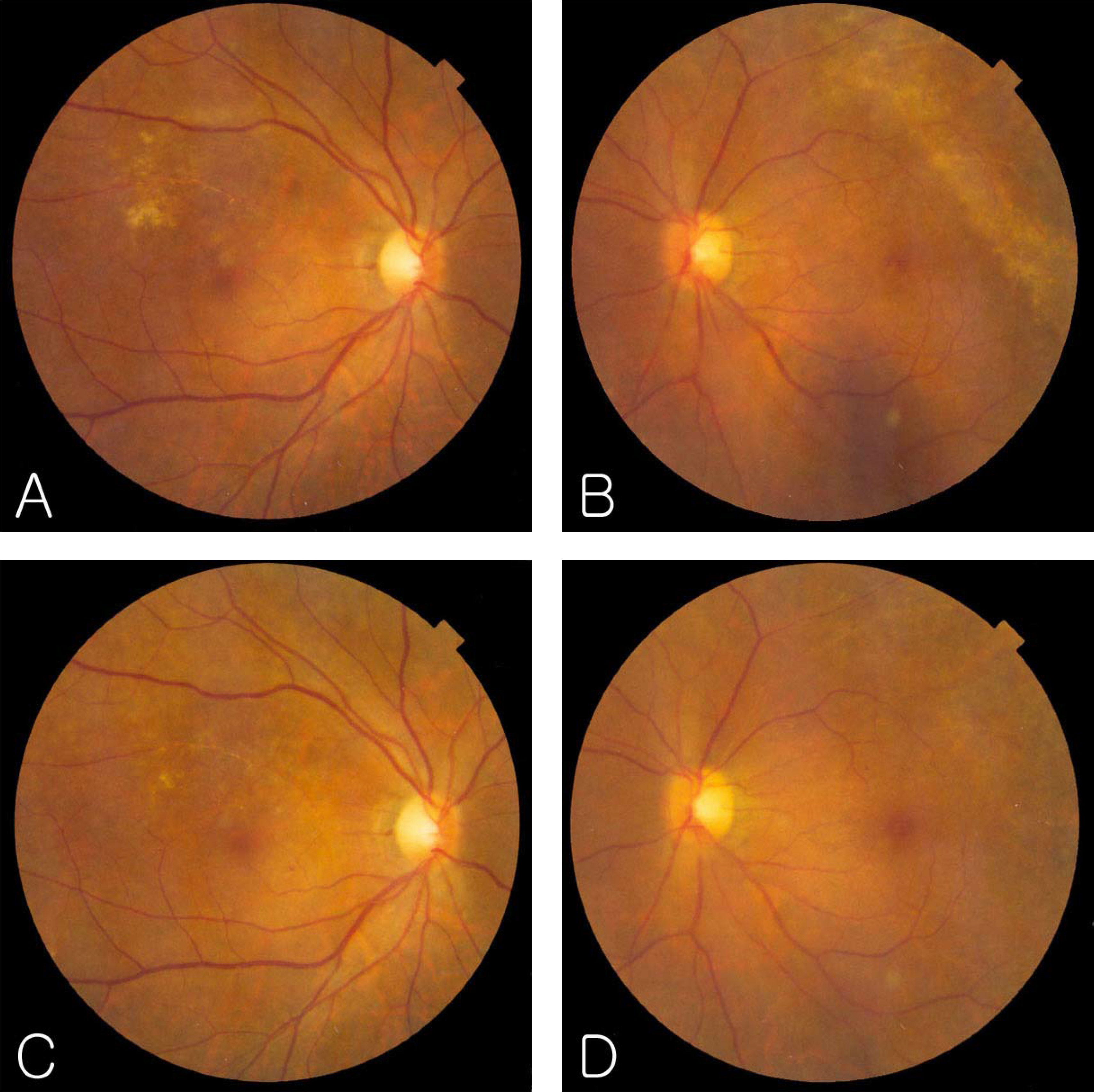
Figure 2. Fundus photographs after intravenous ganciclovir as induction therapy and oral valganciclovir as maintenance therapy.(A, B) After 2 weeks of intravenous ganciclovir as induction therapy, the granular lesions have decreased and the lesion border not advanced. (C, D) After 2 weeks of oral valganciclovir as maintenance therapy, the lesions have much decreased. Retinal vascular sheathing observed at the superotemporal portion of the left eye.

Figure 3. Fundus photographs following reactivation of CMV retinitis. (A, B) CMV retinitis lesion following reactivation at 4 weeks after intravenous ganciclovir as induction therapy and oral valganciclovir as maintenance therapy were discontinued. The lesion border advanced more anteriorly and in circumferential directions than in the first attack. (C, D) After 2 weeks of oral valganciclovir as maintenance therapy, the granular lesions became less opacified. The area of infected retinal tissue has been replaced by a thin, gliotic scar, with the fine mottling of the retinal pigment epithelium.
Cited by 1 articles
-
Cinical Manifestations and Prognosis of Cytomegalovirus Retinitis
Young Kyo Kwun, Ju Byung Chae, Don Il Ham
J Korean Ophthalmol Soc. 2010;51(2):203-209. doi: 10.3341/jkos.2010.51.2.203.
Reference
-
References
1. Egbert PR, Pollard Rb, Gallagher JG, et al. Cytomegalovirus retinitis in immunosuppressed hosts. II. Ocular manifestations. Ann Intern Med. 1980; 93:664–70.2. Pescoviz MD, Rabkin J, Merion RM, et al. Valganciclovir results in improved oral absorption of ganciclovir in liver transplant recipients. Antimicrob Agents Chemother. 2000; 44:2811–5.
Article3. Limaye AP, Corey L, Koelle DM, et al. Emergence of ganciclovir-resistant cytomegalovirus disease among recipients of solid-organ transplants. Lancet. 2000; 356:645–9.
Article4. Markham A, Faulds D. Ganciclovir. An update of its therapeutic use in cytomegalovirus infection. Drugs. 1994; 48:455–84.5. Pescovitz MD, Pruett TI, Gonwa T, et al. Oral ganciclovir dosing in transplant recipients and dialysis patients based on renal function. Transplantation. 1998; 66:1104–7.6. Bartlett JG. The Johns Hopkins Hospital 1997 guide to medical care of patients with HIV infection. 7th ed.Baltimore: Williams & Wilkins;1997. p. 107–9.7. Einsele H, Reusser P, Bornhauser M, et al. Oral valganciclovir leads to higher exposure to ganciclovir than intravenous ganciclovir in patients following allogeneic stem cell transplantation. Blood. 2006; 107:3002–8.
Article8. Faulds D, Heel RC. Ganciclovir. A review of its antiviral activity, pharmacokinetic properties and therapeutic efficacy in cytomegalovirus infections. Drugs. 1994; 48:455–84.9. Hamzeh FM, Literman PS. Intranuclear accumulation of sub-genomic noninfectious human cytomegalovirus DNA in infected cells in the presence of ganciclovir. Antimicrob Agents Chemother. 1991; 35:1818–23.
Article10. Jung D, Dorr A. Single-dose pharmacokinetics of valganciclovir in HIV- and CMV-seropositive subjects. J Clin Pharmacol. 1999; 39:800–4.
Article11. Brown F, Banken L, Saywell K, Arum I. Pharmoacokinetics of valganciclovir and ganciclovir following multiple oral dosages of valganciclovir in HIV- and CMV-serppositive volunteers. Clin Pharmacokinet. 1999; 37:167–76.12. Martin DF, Sierra-Madero J, Walmsley S, et al. A controlled trial of valganciclovir as induction therapy for cytomegalovirus retinitis. N Engl J Med. 2002; 346:1119–26.
Article13. Kim IT, Lim JH, Seo HD. Ganciclovir treatment for cytomegalovirus retinitis in renal transplant recipients. J Korean Ophthalmol Soc. 1997; 38:242–50.14. Holland GN, Sidikaro Y, Kreiger AE, et al. Treatment of cytomegalovirus retinopathy with ganciclovir. Ophthalmology. 1987; 94:815–23.
Article15. Jabs DA, Newman C, de Bustros S, et al. Treatment of cytomegalovirus retinitis with ganciclovir. Ophthalmology. 1987; 94:824–30.
Article16. Kim YH, Kim SK. Cytomegalovirus retinitis in a child with acute lymphoblastic leukemia. J Korean Ophthalmol Soc. 2006; 47:1009–15.17. Polland RB, Egbert PR, Gallagher JG, Merigan TC. Cyto-megalovirus retinitis in immunosuppressed hosts. I. Natural history and effects of treatment with adenine arabinoside. Ann Intern Med. 1980; 93:655–64.18. Meredith TA, Aaberg TM, Resser FH. Rhegmatogenous retinal detachment complicating cytomegalovirus retinitis. Am J Ophthalmol. 1979; 87:793–6.
Article19. Erice A, Jordan MC, Chace BA, et al. Ganciclovir treatment of cytomegalovirus disease in transplant recipients and other immunocompromised hosts. JAMA. 1987; 12:3082–7.
Article20. Harbison MA, DeGirolami PC, Jenkins RL, et al. Ganciclovir therapy of severe cytomegalovirus infections in solid organ transplant recipients. Transplantation. 1988; 46:82–8.21. Banmal CR, Levin AV, Read SE, et al. Cytomegalovirus retinitis in immunosupressed children. Am J Ophthalmol. 1999; 127:550–8.22. Park MY, Ohn YH, Park SH. Three cases of cytomegalovirus retinitis in the immunosupressed kidney transplant patients. J Korean Ophthalmol Soc. 1993; 34:918–23.23. Yi WM, Kim MH, Yoo JS, Huh W. CMV papillitis in renal transplant recipient. J Korean Ophthalmol Soc. 1998; 39:2768–71.24. Cho CW, Park YM, Seo MS. Effect of ganciclovir on cytomegalovirus retinitis of a renal transplant patient without maintenance therapy. J Korean Ophthalmol Soc. 1997; 38:637–42.25. Lalezari J, lindely J, Walmsley S, et al. A safety study of oral valganciclovir maintenance treatment of cytomegalovirus retinitis. J Acquir Immune Defic Syndr. 2002; 30:392–400.
Article
- Full Text Links
-
- Actions
-
Cited
- CITED
-
- Close
- Share
-
- Similar articles
-
- Cytomegalovirus Retinitis in a Hematopoietic Stem Cell Transplant Recipient During Maribavir Pre-emptive Therapy
- Therapeutic Effect of Ganciclovir on Cytomegalovirus Retinitis
- Ocular Ischemic Syndrome as the Initial Presenting Feature of Cytomegalovirus Retinitis
- 4 Cases of Reactivated Cytomegalovirus Retinitis in Immunocompromised Patients
- Cytomegalovirus Retinopathy in Aequired Immunodeficieney Syndrome
