A Clinical Analysis of Surgical Treatment of Lumbar Degenerative Kyphosis
- Affiliations
-
- 1Department of Orthopedic Surgery, National Medical Center, Seoul, Korea. eungha@unitel.co.kr
- KMID: 2209737
- DOI: http://doi.org/10.4184/jkss.2001.8.3.210
Abstract
-
STUDY DESIGN: A retrospective study.
OBJECTIVES
To analyse associated preoperative conditions and postoperative causes of sagittal imbalance and to analyze clinical results of surgical treatment of lumbar degenerative kyphosis. SUMMARY OF LITERATURE REVIEW: There have been many controversies and high possibility of unsatisfactory results in surgical treatment of symptomatic degenerative lumbar kyphosis, which was complicated condition usually needed multi-level operation in old age.
MATERIALS AND METHODS
We analyzed 24 patients who complained of long standing stooping as one of main symptoms with radiologically measured lumbar kyphosis and underwent surgical restoration of lumbar lordodsis with long segmental spinal fusion from 1995 to 1999. Mean follow-up was 31.9months(from 24 to 48 months). Operative treatments were posterolateral fusion with pedicular screw(15 cases), anterior and posterior interbody fusion(5 cases), posterior interbody fusion with cage(1 case) and decancellation osteotomy(3 cases). Cases divided into 2 groups(Group A: improved stooping, Group B: recurred stooping) were evaluated by radiological measurement of changes in surgically restored lumbar lordosis correlated with clinical improvement of stooping. Overall clinical results were evaluated according to Kirkaldy-Willis criteria.
RESULTS
The associated conditions of preoperative lumbar kyphosis were recognized as multiple disc degeneration, segmental instability, degenerative vertebral wedging and pseudospondylolisthesis. Post-operative stooping recurred in 5 cases and caused by adjacent kyphosis in 2 cases, loss of correction in 1 case and both in 2 cases. Loss of correction was associated with pseudarthrosis in 1 case, screw loosening in 3 cases and allograft collapse in 2 cases. According to Kirkaldy-Willis, 8 cases of unsatisfactory clinical results consisted of 3 cases of pseudarthrosis out of 19 cases of Gruop A and all cases(5 cases) in group B. Most of correction loss occurred at lower lumbar spine(L3-S1) and was closely related to post-operative sagittal imbalance.
CONCLUSIONS
Maintenance of well corrected lumbar lordosis for sagittal balance and prevention of pseudarthrosis were mandatory for good clinical outcome in surgical treatment of lumbar degenerative kyphosis.
MeSH Terms
Figure
-
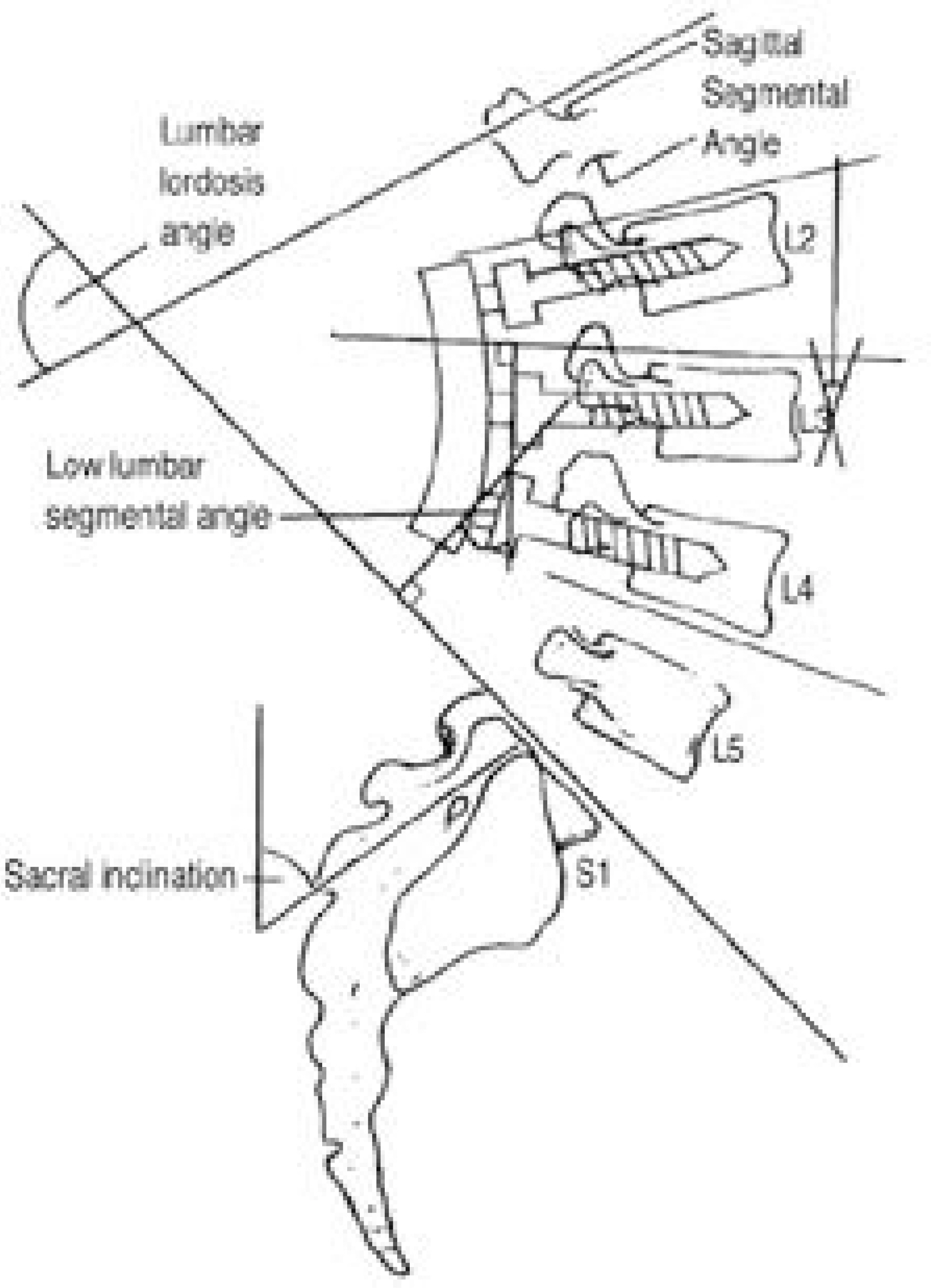
Fig. 1. Parameter which represent spinal alignment.

Fig. 2-A. Preoperative lateral radiography of 68-year-old man shows disc degeneration of L2-3 L3-4 L4-5 L5-S1, and rigid kyphosis in flexion-extension view. Fig. 2-B. Postoperative lateral radiography with L2-S1 posterolateral fusion with pedicular screw and pedicle subtraction osteotomy on L4 shows increased lumbar lordosis angle by 16˚. Postoperative 28-months followup lateral radiography of lumbar spine shows correction loss(L1-S1) by 8˚, but shows maintenance of lumbar lordosis and relief of stooping.
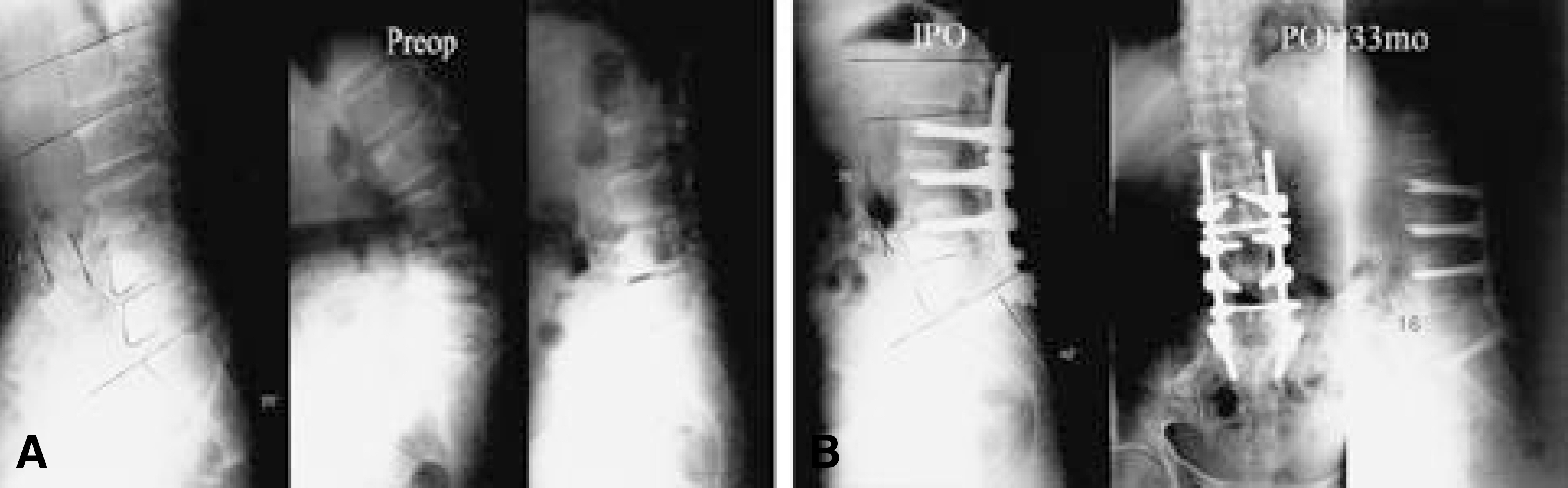
Fig. 3-A. Preoperative lateral radiography of 64-year-old woman shows disc degeneration of L4-5 L5-S1, wedge vertebrae L4 L5, and segmental instability L3-4 L4-5 in flexion-extension view. Fig. 3-B. Postoperative lateral radiography with L2-S1 posterolateral fusion with pedicular screws and L2-5 anterior interbody fusion with allograft shows increased lumbar lordosis angle by 15˚. Postoperative 33-months followup lateral radiography of lumbar spine shows compression fx L1 with correction loss(L1-S1) by 4˚, but showed maintenance of lumbar lordosis and relief of stooping.
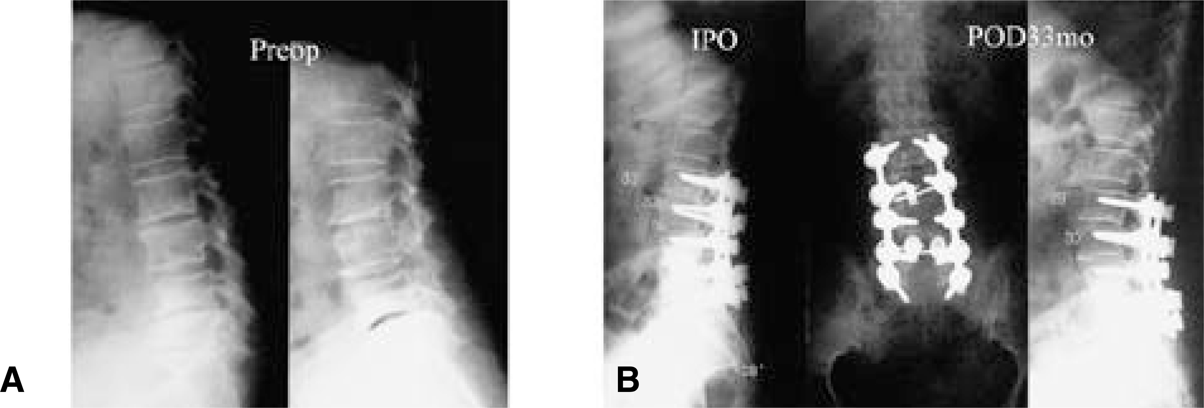
Fig. 4-A. Preoperative lateral radiography of 66-year-old woman shows disc degeneration of L2-3 L4-5 L5-S1, wedge vertebrae L5 and pseudospondylolisthesis L3 on 4. Fig. 4-B. Postoperative lateral radiography with L2-S1 posterolateral fusion with pedicular screw and posterior lumbar interbody fusion with cage L4-5 shows increased lumbar lordosis angle by 11˚. Postoperative 33-months followup lateral radiography of lumbar spine shows correction loss(L1-S1) by 4˚, but showed maintenance of lumbar lordosis and relief of stooping.
Cited by 4 articles
-
Loss of Sagittal Balance and Clinical Outcomes following Corrective Osteotomy for Lumbar Degenerative Kyphosis
Ki-Tack Kim, Sang Hun Lee, Kyung-Soo Suk, Jung-Hee Lee, Yang-Sun Im, Eun-Min Seo
J Korean Orthop Assoc. 2009;44(1):83-92. doi: 10.4055/jkoa.2009.44.1.83.Anterior and Posterior Surgical Treatment with Wedged Cage (SynCageⓇ) in Lumbar Degenerative Kyphosis
Eung-Ha Kim, Sun-Woo Kim
J Korean Soc Spine Surg. 2003;10(3):240-247. doi: 10.4184/jkss.2003.10.3.240.Comparative Analysis of Surgical Options in the Treatment of Lumbar Degenerative Kyphosis
Jae Chul Lee, Jae-Wan Soh, Joo-Hyoung Jo, Yon-Il Kim, Byung-Joon Shin
J Korean Soc Spine Surg. 2009;16(1):8-16. doi: 10.7469/JKSS.2009.16.1.8.Natural History of Lumbar Degenerative Kyphosis with Conservative Treatment
Whoan Jeang Kim, Shann Haw Chang, Gyu Sang Lee, Yong Ho Kim, Kun Young Park, Kyung Hoon Park, Won Sik Choy
J Korean Soc Spine Surg. 2017;24(1):24-31. doi: 10.4184/jkss.2017.24.1.24.
Reference
-
1). Aaro S, Ohlen G. The effect of Harrington instrumentation on the sagittal configuration and mobility of the spine in scoliosis. Spine. 8(6):570–575. 1983.
Article2). Andersson BJG, Ortengren R. Myoelectric back muscle activity during sitting. Scand J Rehab Med. 3(Suppl):73–90. 1974.3). Atsuta Y. Degenerative kyphosis in advanced age; Cause and management(in Japanese). J Jpn Orthop Assoc, 37-. 3:289–295. 1994.4). Becker's L, Bekaert J. The role of lordosis. Acta Orthop Belg. 57(Suppl):198–202. 1991.5). Bernhardt M, Bridwell KH. Segmental analysis of the sagittal plane alignment of normal thoracic and lumbar spines and thoracolumbar junction. Spine. 14:717–721. 1989.6). Chernukha KV, Daffner RH, Reigel DH. Lumbar lordosis measurement. Spine. 23:74–80. 1998.
Article7). Deniel E, Geib MD, Lawrence G, Lenke MD, Keith H, Bridwell MD, Kathy Blanke RN, Kevin W, McBn-ery MD. An-Analysis of Sagittal Spinal Alignment in 100 Asymptomatic Middle and Older Aged Volunteers. Spine. 20:1351–1358. 1995.8). Doherty JH. Complication of fusion in lumbar scoliosis. J Bone Joint Surg. 55-A:438. 1973.9). Fernand R, Fox DE. Evaluation of lumbar lordosis. Spine. 10:799–803. 1985.
Article10). Grobler LJ, Moe JH, Winter RB, et al. Loss of lumbar lordosis following surgical correction of thoracolumbar deformity, Orthop Trans. 2(2):39. 1978.11). Hadar H, Godoth N, Heifetz M. Fatty replacement of lower paraspinal muscles, normal and neuromuscular dis -oders. AJR. 141:895–898. 1983.12). Kirkaldy-Willis WH, Paine KWE, Cauchoix J, Mcl-vor G. Lumbar Spinal Fusion: A biomechanical study, Spine. 9:574–581. 1984.13). Lee CS, Kim YT, Kim EG. Clinical Study of Lumbar Degenerative Kyphosis. J Korean Spine Surg. 4(1):27–35. 1997.14). Lee JS, Oh WH, Chung SS, Lee SG, Lee JY. Analysis of the Sagittal Alignment of Normal Spines. J Korean Orthop. 34:949–954. 1999.
Article15). Michael OLG. Loss of lumbar lordosis: A complication of spinal fusion for scoliosis. Orthop. Clin. N AM, 19-. 2:383–393. 1988.16). Moe JH, Denis F. The iatrogenic loss of lumbar lordosis. Orthop Trans. 1-2:131. 1977.17). Stagnara P, De Mauroy JC, Dran G, et al. Reciprocal angulation of vertebral bodies in sagittal plane: Approach to references in the evaluation of kyphosis and lordosis. Spine. 7:335–342. 1982.18). Takemitsu Y, Harada Y, Iwahara T, Miyamoto M, Mitatake Y. Lumbar Degenerative Kyphosis: Clinical, radiologic and epidemiological study. Spine. 13:1317–1326. 1988.19). Takemitsu Y, Harada Y, Iwahara T. Low back pain and aging change of spine in Japanese farmers aged more than 40 years. J Jpn Orthop Assoc. 58:551–552. 1984.20). Wambolt A, Spencer DL. A segmental analysis of the distribution of lumbar lordosis in the normal spine. Orthop Trans. 11:92–93. 1987.21). Winter RB. Harrington instrumentation into the lumbar spine, technique for preservation of normal lumbar lordosis. Spine. 11(9):633–635. 1986.22). Yohichi Aota, Kiy oshi Kumano, and Shigeru Hiraba-yashi. Postfusion instability at the adjacent Segments after rigid pedicle screw fixation for degenerative lumbar spinal disorders. Journal of Spinal Disorders. 8(6):464–473. 1995.
- Full Text Links
-
- Actions
-
Cited
- CITED
-
- Close
- Share
-
- Similar articles
-
- Clinical Study of Lumbar Degenerative Kyphosis
- Classification and Treatment of Degenerative Lumbar Spinal Deformity
- Proximal Junctional Problems in Surgical Treatment of Lumbar Degenerative Sagittal Imbalance Patients and Relevant Risk Factors
- Comparative Analysis of Surgical Options in the Treatment of Lumbar Degenerative Kyphosis
- Comparative Radiographic Outcomes of Lateral and Posterior Lumbar Interbody Fusion in the Treatment of Degenerative Lumbar Kyphosis
