Multiple Extradural Arachnoid Cyst : A Case Report
- Affiliations
-
- 1Department of Orthopedic Surgery, Seoul Medical Center, Seoul, Korea. hananina@dreamwiz.com
- KMID: 2209606
- DOI: http://doi.org/10.4184/jkss.2009.16.2.122
Abstract
- Multiple extradural arachnoid cysts of the spine are extremely uncommon in children with only a few cases reported. The authors report a case of multiple extradural spinal arachnoid cysts in children with a review of the relevant literature.
Keyword
MeSH Terms
Figure
-
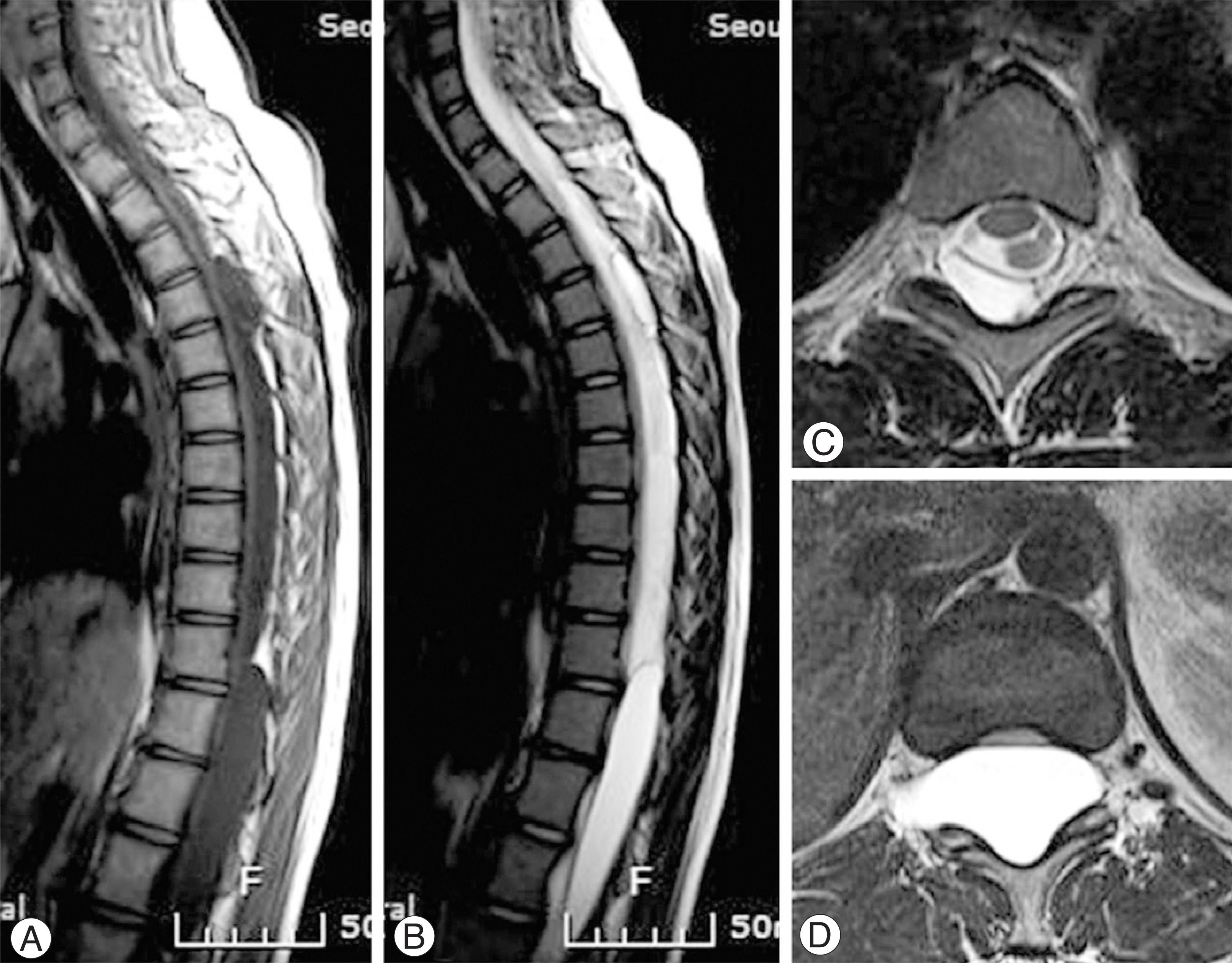
Fig. 1. Preoperative sagittal T1-weighted (A) and T2-weighted (B) spine MR images demonstrating extensive cystic lesion causing spinal cord compression. The spinal cord is pushed ventrally in the spinal canal. Axial T2-weighted MR images obtained at T4 (C) and T11 (D), showing that the cystic lesions are extradural in location.

Fig. 2. Intraoperative photographs showing spinal arachnoid cysts. A T10-L2 laminectomy was performed to expose the large extradural spinal arachnoid cyst (A) and thecal sac can be identified to allow dissection of the cyst (B).
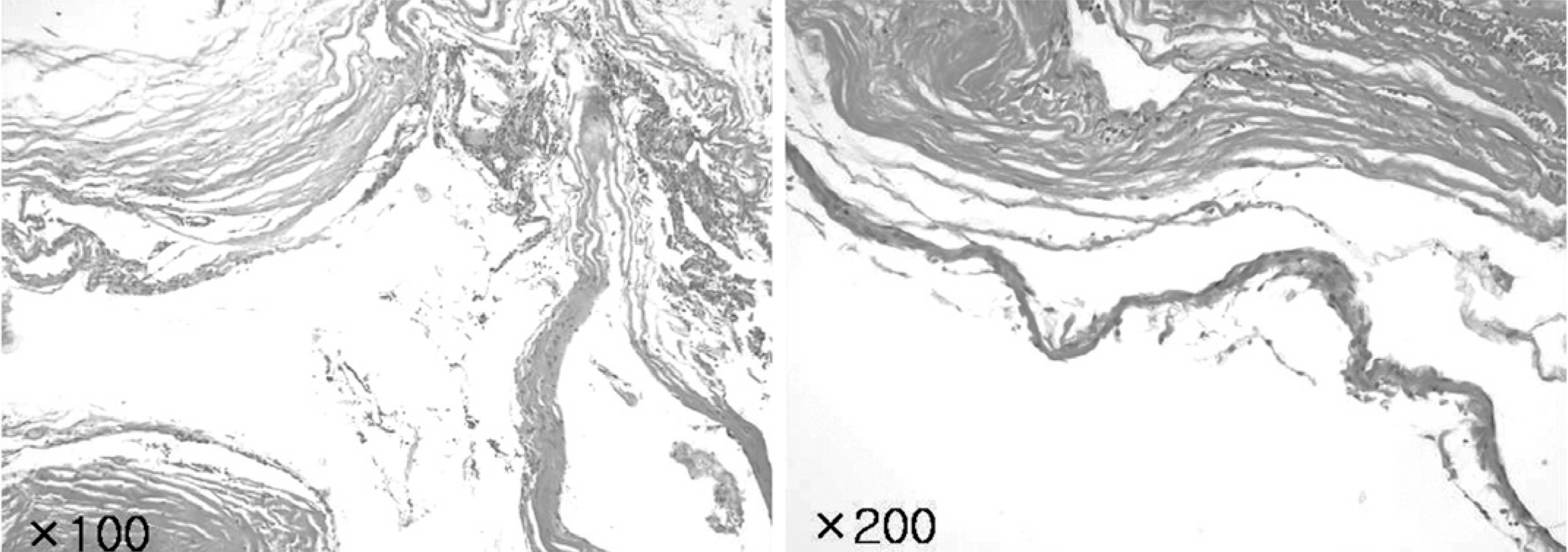
Fig. 3. Photomicrograph of cyst wall demonstrating layered collagenous fibers and epithelial tissue with flat lining cells. H & E, ×100, 200.
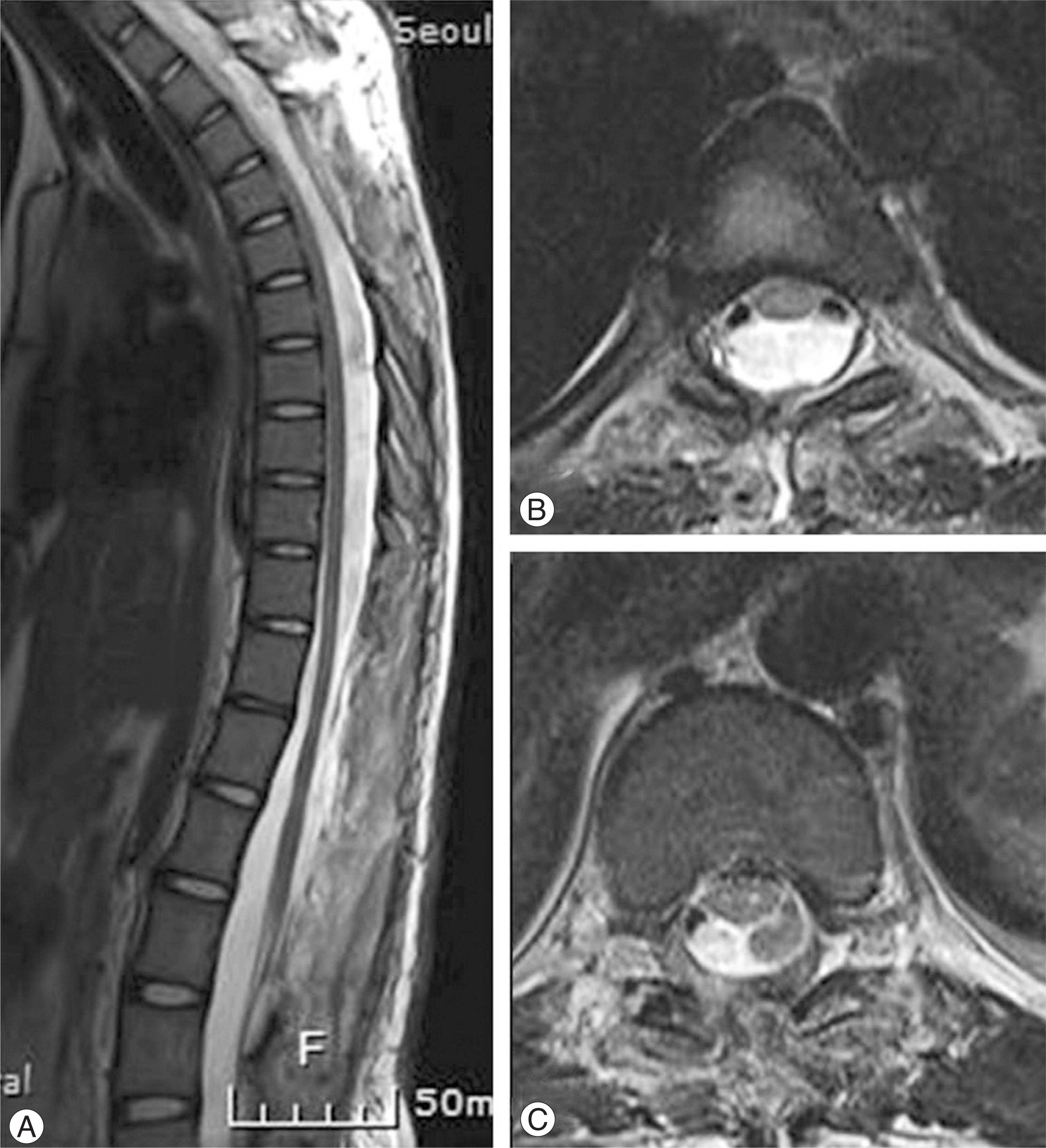
Fig. 4. Sagittal T2-weighted (A) and Axial T2-weighted (B, C) MR images obtained 2 weeks postoperatively demonstrating expanded spinal cord, complete excision of multiple extradural arachnoid cysts.
Reference
-
1). Erisberg CA, Dyke CG, Brewer DE. Symptoms and diagnosis of extradural cysts. Bull Neurol Inst NY. 1934; 3:395–417.2). Rabb C, McComb J, Raffel C, Kennedy J. Spinal arachnoid cyst in the pediatric age group: an association with neural tube defect. J Neurosurg. 1992; 77:369–372.3). Spiller WG. A case of intradural spinal cyst with operation and recovery. Trans Coll Physicians Philadelphia. 1903; 25:1–18.4). Cilluffo JM, Gomez MR, Reese DF, Onofrio BM, Miller RH. Idiopathic (“congenital”) spinal arachnoid diverticula. Clinical diagnosis and surgical results. Myo Clin Proc. 1981; 56:93–101.5). Nabors MW, Pait TG, Byrd EB, Karim NO, Davis DO, Kobrine AI. Updated assessment and current classification of spinal meningeal cysts. J Neurosurg. 1988; 68:366–377.
Article6). Liu JK, Cole CD, Kan P, Schmidt MH. Spinal extradural arachnoid cysts: clinical, radiological, and surgical features. Neurosurg Focus. 2007; 22:6.
Article7). DiSclafani A 2nd, Canale DJ. Communicating spinal arachnoid cysts: diagnosis by delayed metrizamide computed tomography. Surg Neurol. 1985; 23:428–30.8). Fortuna A, La Torre E, Ciappetta P. Arachnoidal diverticula: a unitary approach to spinal cysts communicating with the cubarachnoid space. Acta Neurochir. 1977; 39:259–268.9). Bergland RM. Congenital intraspinal extradural syst. Report of three cases in one family. J Neurosurg. 1968; 28:495–499.10). Ersahin Y, Yildizhan A, Seber N. Spinal extradural arachnoid cyst. Childs Nerv Syst. 1993; 9:250–252.
Article11). Stechison MT, Hendrick EB, Cohen E. Spinal extradural arachnoid cyst: Pediatr Neurosci. 1989; 15:36–38.
- Full Text Links
-
- Actions
-
Cited
- CITED
-
- Close
- Share
-
- Similar articles
-
- Retroperitoneal Hematoma after Excision of Lumbar Extradural Arachnoid Cyst: Case Report
- Extradural Spinal Arachnoid Cyst as a Cause of Cauda Equina Syndrome in a Child
- Spinal Extradural Arachnoid Cyst: Minimally Invasive Surgical Treatment after Localization of Dural Defect Using Magnetic Resonance Myelogram
- Septated Extradural Arachnoid Cyst in Thoracolumbar Spine Causing Myelopathy
- Huge Thoracolumbar Extradural Arachnoid Cyst
