External Snapping Hip Syndrome: Emphasis on the MR Imaging
- Affiliations
-
- 1Department of Radiology, The Catholic University of Korea, St. Paul's Hospital, Seoul, Korea.
- 2Department of Radiology, The Catholic University of Korea, Bucheon St. Mary's Hospital, Bucheon, Korea. mssung99@catholic.ac.kr
- 3Department of Orthopedic Surgery, The Catholic University of Korea, Bucheon St. Mary's Hospital, Bucheon, Korea.
- 4Department of Radiology, The Catholic University of Korea, St. Mary's Hospital, Seoul, Korea.
- 5Department of Radiology, The Catholic University of Korea, St. Vincent's Hospital, Suwon, Korea.
- KMID: 2208968
- DOI: http://doi.org/10.3348/jksr.2010.62.2.185
Abstract
-
PURPOSE: The aim of this study is to evaluate the MR imaging features of patients with external snapping hip syndrome.
MATERIALS AND METHODS
We retrospectively reviewed 63 hip MR images. The images were analyzed according to the thickness and contour of the iliotibial band and the gluteus maximus, the presence of bone marrow edema, bursitis, joint effusion and other associated findings.
RESULTS
The MR imaging of 22 hips with snapping hip syndrome depicted the causes of external snapping hip syndrome in twenty cases (90%). The MR imaging features of the snapping hip included thickening of the iliotibial band in twelve cases (55%) and/or thickening of the anterior band of the gluteus maximus in nineteen (86%), and a wavy contour of the iliotibial band or the anterior band of the gluteus maximus in ten cases (45%). These findings show a significant p value (<0.01).
CONCLUSION
The majority of patients with snapping hip syndrome revealed thickening of the iliotibial band, thickening of the anterior band of the gluteus maximus and wavy contour of the those structures on MR imaging.
MeSH Terms
Figure
-
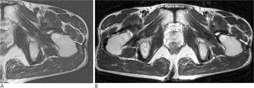
Fig. 1 A 56-years-old man with left snapping hip syndrome. Marked thickening of the iliotibial band (arrow) can be nicely noted on the T2-weighted MR images with a small FOV (180 mm) (A) and a large FOV (330 mm) (B), as compared with the iliotibial band of the right hip.

Fig. 2 A-24-years old man with snapping hip syndrome of both hips. The anterior border of the gluteus maximus of the right hip is thickened (arrow) (A). The left iliotibial band (double arrow) is thicker than that of right iliotibial band on the T2-weighted MR images (B).
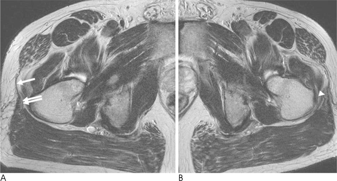
Fig. 3 A 48-years-old man with left snapping hip syndrome. (A) Note the normal finding of the right iliotibial band (arrow) on the axial T2-weighted image. The 2 musculotendinous attachments of the iliotibial band consist of the anterior tensor fascia lata and the posterior gluteus maximus (double arrow). (B) The T2-weighted MR images (FOV 180 mm) show thickening of the left anterior border of the gluteus maximus (arrowhead).
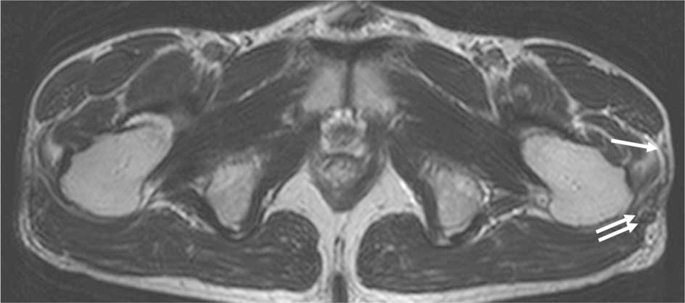
Fig. 4 A 38-years-old man with left snapping hip syndrome. The initial T2-weighted axial MR image of the left hip did not give the any information about the left snapping hip. However, two weeks later, reexamination of both hips with the T2-weighted axial images shows a wavy contour of the iliotibial band (arrow) and irregularity of the anterior border of the gluteus maximus (double arrow) of the left affected hip, as compared with that of the unaffected right hip.

Fig. 5 A 44-years-old man with bilateral snapping hip syndrome. In addition to the iliotibial band lesion (it is not shown best on this section), fibrosis of intermediate signal intensity (arrow) on the axial T2-weighted image (A) and increased signal intensity (arrow) on the fat suppressed T2-weighted images (B) can be seen in the space between the greater trochanter and the iliotibial band.
Reference
-
1. Allen WC, Cope R. Coxa saltans. The snapping hip revisited. J Am Acad Orthop Surg. 1995; 3:303–308.2. Staple TW, Jung D, Mork A. Snapping tendon syndrome: hip tenography with fluoroscopic monitoring. Radiology. 1998; 166:873–874.3. Pelsser V, Cardinal E, Hobden R, Aubin B, Lafortune M. Extraarticular snapping hip: sonographic Findings. AJR Am J Roentgenol. 2001; 176:67–73.4. Choi YS, Lee SM, Song BY, Paik SH, Yoon YK. Dynamic sonography of external snapping hip syndrome. J Ultrasound Med. 2002; 21:753–758.5. Krishnamurthy G, Connolly BL, Narayanan U, Babyn PS. Imaging findings in external snapping hip syndrome. Pediatr Radiol. 2007; 37:1272–1274.6. Faraj AA, Moulton A, Sirivastava VM. Snapping iliotibial band: report of ten cases and review of the literature. Acta Orthop Belg. 2001; 67:19–23.7. Larsen E, Johansen J. Snapping hip. Acta Orthop Scand. 1986; 57:168–170.8. Mayer L. Snapping hip. Surg Gynecol Obstet. 1919; 29:425–429.9. Brignall CG, Brown RM, Stainsby GD. Fibrosis of the gluteus maximus as a cause of snapping hip. J Bone Joint Surg Am. 1993; 75:909–910.10. Schaberg JE, Harper MC, Allen WC. The snapping hip syndrome. Am J Sports Med. 1984; 12:361–365.11. Zoltan DJ, Clancy WG JR, keene JS. A new operative approach to snapping hip and refractory trochanteric bursitis in athletes. Am J Sports Med. 1986; 14:201–204.
- Full Text Links
-
- Actions
-
Cited
- CITED
-
- Close
- Share
-
- Similar articles
-
- Osteochondroma Arising from Anterior Inferior Iliac Spine as a Cause of Snapping Hip
- Snapping Knee caused by the Gracilis and Semitendinosus tendon
- Misdiagnosed Snapping Triceps Syndrome on Ulnar Nerve Dislocation
- Arthroscopic Treatment for External Snapping Hip
- External Snapping Hip Treated by Effective Designed N-plasty of the Iliotibial Band
