Development of a Korean Standard Structural Brain Template in Cognitive Normals and Patients with Mild Cognitive Impairment and Alzheimer's Disease
- Affiliations
-
- 1Department of Biomedical Engineering, Kyunghee University, Youngin, Korea.
- 2Department of Radiology, Kyunghee University Hospital-Gangdong, School of Medicine, Kyunghee University, Seoul, Korea. ghjahng@gmail.com
- 3Department of Neurology, Gangdong Kyunghee University Hospital-Gangdong, School of Medicine, Kyunghee University, Seoul, Korea.
- KMID: 2206907
- DOI: http://doi.org/10.13104/jksmrm.2010.14.2.103
Abstract
- PURPOSE
To generate a Korean specific brain template, especially in patients with Alzheimer's disease (AD) by optimizing the voxel-based analysis.
MATERIALS AND METHODS
Three-dimensional T1-weighted images were obtained from 123 subjects who were 43 cognitively normal subjects and patients with 44 mild cognitive impairment (MCI) and 36 AD. The template and the corresponding aprior maps were created by using the matched pairs approach with considering differences of age, gender and differential diagnosis (DDX). We measured several characteristics in both our and the MNI templates, including in the ventricle size. Also, the fractions of gray matter and white matter voxels normalized by the total intracranial were evaluated.
RESULTS
The high resolution template and the corresponding aprior maps of gray matter, white matter (WM) and CSF were created with the voxel-size of 1 x 1 x 1 mm. Mean distance measures and the ventricle sizes differed between two templates. Our brain template had less gray matter and white matter areas than the MNI template. There were volume differences more in gray matter than in white matter.
CONCLUSION
Gray matter and/or white matter integrity studies in populations of Korean elderly and patients with AD are needed to investigate with this template.
Keyword
Figure
-

Fig. 1 Flowchart for the brain template creation. In this study, we use two separated steps, VBM5 and TOM. GM: Gray matter WM: White matter CSF: Cerebrospinal Fluid VBM: Voxel-Based Morphometry TOM: Template-O-Matic

Fig. 2 Measured parameters shown on the images of the smoothed our brain template (a, c) and the MNI-152 template (b, d). The solid lines show the center position (0, 0, 0) in x, y z plane. The upper and lower dotted lines show the length of the SP-IP size and the middle dotted line is the length of the AP-PP line. The vertical lines show the length of the RP-LP line.
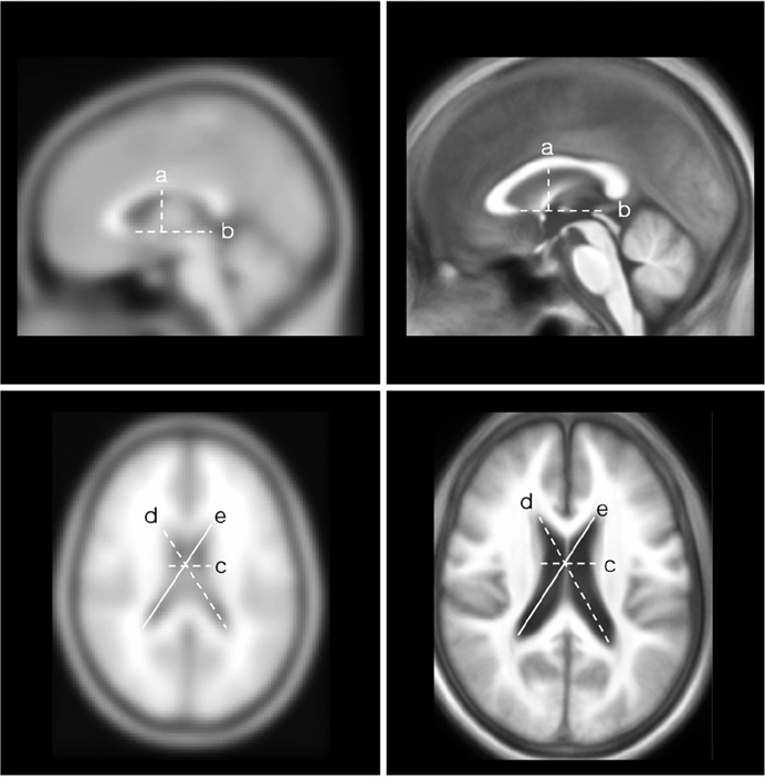
Fig. 3 Measured distances (mm) of the ventricle area for selecting landmark sites in the MNI-152 template (Upper left and bottom left) and the smoothed our brain template (Upper right and bottom right).
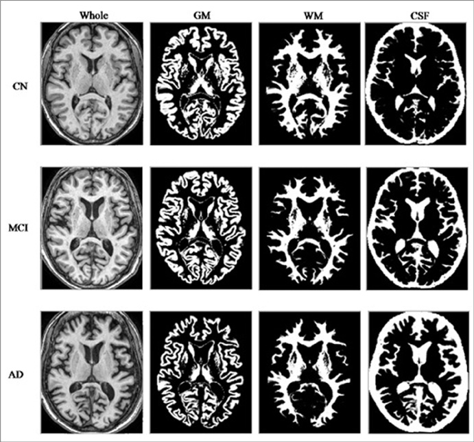
Fig. 4 Representative segmented images obtained in a cognitively normal (CN) control subject (upper row), in a patient with mild cognitive impairment (MCI, middle row), and in a patient with Alzheimer's disease (AD, bottom row). Each subject is a 70 years-old woman. GM: Gray matter WM: White matter CSF: Cerebrospinal Fluid

Fig. 5 The created standard brain templates of three-dimensional T1-weighted (a) and the corresponding tissue maps of gray matter(GM, b), white matter (WM, c), andcerebrospinal fluid (CSF, d).
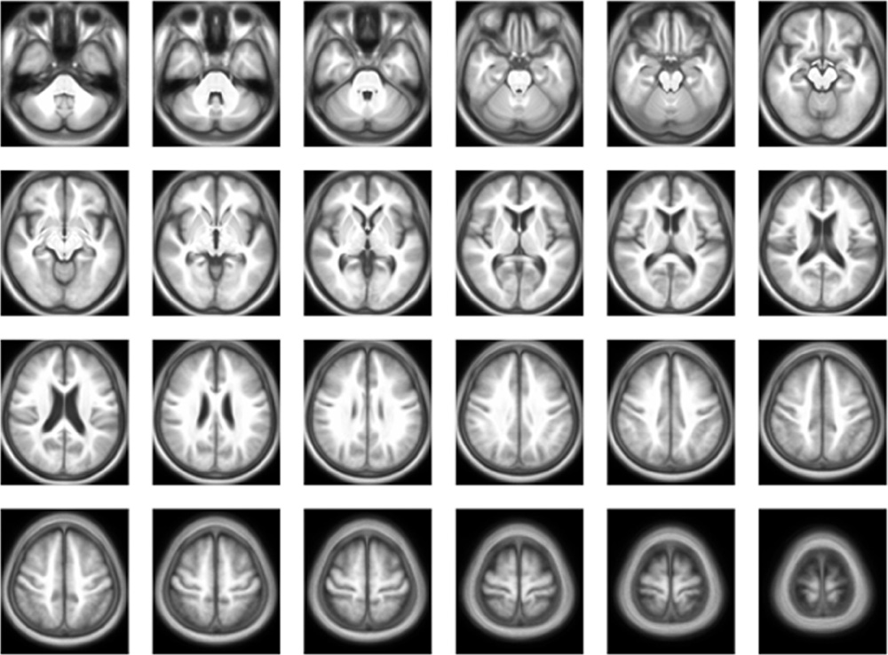
Fig. 6 The created standard brain template of three-dimensional T1-weighted images was shown as the axial plane slices.

Fig. 7 Beta image volumes in a general linear model; The ages are the coefficients of the third order polynomial Maps from the second row to the sixth row represent beta image volumes in a general linear model of the standard templates caused by the co-varietiesof age, gender, and DDX. GM: Gray matter WM: White matter CSF: Cerebrospinal Fluid DDX: Differential diagnosis
Cited by 1 articles
-
Voxel-based Investigations of Phase Mask Effects on Susceptibility Weighted Images
Eo Jin Hwang, Min Ji Kim, Hyug Gi Kim, Chang Woo Ryu, Geon Ho Jahng
Prog Med Phys. 2013;24(1):25-34. doi: 10.14316/pmp.2013.24.1.25.
Reference
-
1. Brant-Zawadzki M, Gillan GD, Nitz WR. MP RAGE: a three-dimensional, T1-weighted, gradient-echo sequence--initial experience in the brain. Radiology. 1992. 182:769–775.2. Yamashita E, et al. Evaluation of three-dimensional fast spoiled gradient recalled acquisition in the steady state (FSPGR) using ultra magnetic field 3-Tesla MRI for optimal pulse sequences of T1-weighted imaging. Nihon Hoshasen Gijutsu Gakkai Zasshi. 2006. 62:297–304.3. Pruessner JC, et al. Volumetry of hippocampus and amygdala with high-resolution MRI and three-dimensional analysis software: minimizing the discrepancies between laboratories. Cereb Cortex. 2000. 10:433–442.4. Ashburner J, Friston KJ. Voxel-based morphometry--the methods. Neuroimage. 2000. 11:805–821.5. Evans AC, et al. Anatomical mapping of functional activation in stereotactic coordinate space. Neuroimage. 1992. 1:43–53.6. Braak H, Braak E. Neuropathological stageing of Alzheimer-related changes. Acta Neuropathol. 1991. 82:239–259.7. Lim HK, Choi EH, Lee CH. A voxel-Based Morphometry of gray matter reduction in patients with dementia of the Alzheimer's type. Korean J Biol Psychiatry. 2008. 15:118–125.8. Kakeda S, Korogi Y. The efficacy of a voxel-based morphometry on the analysis of imaging in schizophrenia, temporal lobe epilepsy, and Alzheimer's disease/mild cognitive impairment: a review. Neuroradiology. 2010.9. Frisoni GB, et al. The Clinical Use of Structural MRI in Alzheimer Disease. Nat Rev Neurol. 2010. 6:67–77.10. Choi SH, et al. Optimized VBM in patients with alzheimer's disease: gray matter loss and its correlation with cognitive function. J Korean Radiol Soc. 2005. 53:323–329.11. Abe O, et al. Voxel-based analysis of the diffusion tensor. Neuroradiology. 2010.12. Jahng GH, Schuff N. Influence of selecting EPI readout-encoding bandwidths on arterial spin labeling perfusion MRI. MAGMA. 2009. 22:287–295.13. Schweinhardt P, et al. A template for spatial normalisation of MR images of the rat brain. J Neurosci Methods. 2003. 129:105–113.14. Tang S, et al. RABBIT: rapid alignment of brains by building intermediate templates. Neuroimage. 2009. 47:1277–1287.15. Kazemi K, et al. A neonatal atlas template for spatial normalization of whole-brain magnetic resonance images of newborns: preliminary results. Neuroimage. 2007. 37:463–473.16. Choi DY, et al. Development of Korean standard brain templates according to gender and age. Korean J Anat. 2004. 37:255–261.17. Lee JS, et al. Development of Korean standard brain templates. J Korean Med Sci. 2005. 20:483–488.18. Shin DH, et al. A study on the deviation of cluster based on template images of Korean childrens brain SPECT image using the statistical parametic mapping. Korean J Med Phys. 2004. 15:45–53.19. Talairach J, Tournoux P. Co-planar stereotaxic atlas of the human brain: 3-dimensional proportional system-an approach to cerebral imaging. 1988. Thieme Medical Publishers;1.20. Lancaster JL, et al. Bias between MNI and Talairach coordinates analyzed using the ICBM-152 brain template. Hum Brain Mapp. 2007. 28:1194–1205.21. Zilles K, et al. Hemispheric shape of European and Japanese brains: 3-D MRI analysis of intersubject variability, ethnical, and gender differences. Neuroimage. 2001. 13:262–271.22. Chung SC, et al. The voumetric study of the ventricle in Korean according to age and gender. Korean J Anat. 2005. 38:207–213.23. May A, Gaser C. Magnetic resonance-based morphometry: a window into structural plasticity of the brain. Curr Opin Neurol. 2006. 19:407–411.24. Wells WM, et al. Adaptive segmentation of MRI data. IEEE Trans Med Imaging. 1996. 15:429–442.25. Wilke M, et al. Template-O-Matic: a toolbox for creating customized pediatric templates. Neuroimage. 2008. 41:903–913.26. Franke K, et al. BrainAGE: a completely automated age estimation framework using structural MRI. 2010. In : 16th Annual Meeting of the Organization for Human Brain Mapping; Barcelona, Spain.27. Karas G, et al. Precuneus atrophy in early-onset Alzheimer's disease: a morphometric structural MRI study. Neuroradiology. 2007. 49:967–976.28. Lee JS, et al. Development of Korean Standard Brain Templates. J Korean Med Sci. 2005. 20:483–488.
- Full Text Links
-
- Actions
-
Cited
- CITED
-
- Close
- Share
-
- Similar articles
-
- The Clinical Significance of Cognitive Interventions for the Patients with Mild Cognitive Impairment
- Neuroimaging Markers for Alzheimer's Disease and Mild Cognitive Impairment in Alzheimer's Disease Neuroimaging Initiative (ADNI)
- Cognitive Assessment in Traumatic Brain Injury
- Mild Cognitive Impairment - Aging to Alzheimer's Disease -
- Subtypes and Treatment of Mild Cognitive Impairment
