Micronodular Thymoma with Lymphoid Stroma: A Case Report
- Affiliations
-
- 1Department of Pathology, Sungkyunkwan University School of Medicine, Seoul, Korea. hanjho@skku.edu
- 2Department of Radiology, Sungkyunkwan University School of Medicine, Seoul, Korea.
- KMID: 2199896
- DOI: http://doi.org/10.6058/jlc.2011.10.1.56
Abstract
- Micronodular thymoma with lymphoid stroma (MNT) is an extremely rare tumor and has not been reported in Korea. Herein, we report a case of MNT diagnosed in a 75 year-old male. The mass was incidentally identified on thoracic computer tomography (CT) during work-up for a cardiac mass. The resected thymic mass was ovoid and solid measuring 3.5x3 cm in size and showed homogenously white tan, solid and firm cut surface. Microscopically, the tumor had a thin fibrous capsule except small foci of invasion to thymic adipose tissue, and consisted of characteristic multiple discrete epithelial nodules in abundant lymphoid stroma. The epithelial nests anastomosed each other, forming a vague cord-like structure. The cells in the epithelial nests were bland, spindle to oval shape with relatively abundant cytoplasm, slightly vesicular chromatin and one definite nucleolus. The lymphoid stroma contained prominent germinal centers. No evidence of recurrence or metastasis has been found within 4 years of surgery.
Keyword
MeSH Terms
Figure
-
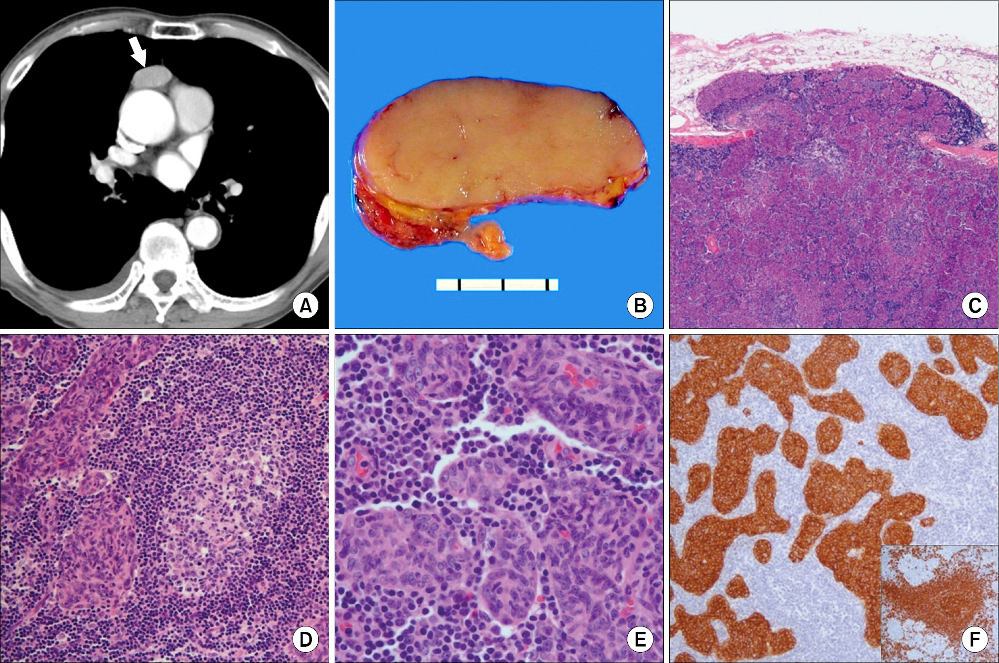
Fig. 1. (A) A well-demarcated mass (arrow) was found in the anterior mediastinum on thoracic computer tomography (CT). (B) Cut section of the mass was solid and lobulated with fine fibrous septa. (C) The tumor showed focal extension beyond the capsule to peri-thymic adipose tissue (hematoxylin-eosin stain, ×40). (D) Multiple lymphoid follicles with germinal centers were found in the lymphoid stroma (hematoxylin-eosin stain, ×200). (E) Discrete epithelial nodules in lymphoid-rich stroma were unique microscopic feature (hematoxylin-eosin stain, ×400). (F) Cytokeratin 5/6 was positive in the epithelial components, and CD 20 was positive in lymphoid stromal cells (inset) (ABC method, ×100).
Cited by 1 articles
-
A Rare Case of Mixed Type A Thymoma and Micronodular Thymoma with Lymphoid Stroma
Yoon Jin Cha, Joungho Han, Jimin Kim, Kyung Soo Lee, Young Mog Shim
J Pathol Transl Med. 2015;49(1):75-77. doi: 10.4132/jptm.2014.10.27.
Reference
-
1. Travis WD, Brambilla E, Muller-Hermelink HK, Harris CC, editors. World Health Organization Classification of Tumours. Pathology and genetics of tumours of the lung, pleura, thymus and heart. Lyon: IARC Press;2004. p. 148–151.2. Suster S, Moran CA. Micronodular thymoma with lymphoid B-cell hyperplasia: clinicopathologic and immunohistochemi-cal study of eighteen cases of a distinctive morphologic variant of thymic epithelial neoplasm. Am J Surg Pathol. 1999; 23:955–962.3. El MF, Braham E, Ayadi A, Ismail O, Kilani T. Micronodular thymoma with lymphoid troma: report of two cases and particular association with thymic lymphoid hyperplasia in one case. Pathology. 2006; 38:586–588.4. Tateyama H, Saito Y, Fujii Y, et al. The spectrum of micronodular thymic epithelial tumours with lymphoid B-cell hyperplasia. Histopathology. 2001; 38:519–527.
Article
- Full Text Links
-
- Actions
-
Cited
- CITED
-
- Close
- Share
-
- Similar articles
-
- Micronodular Thymoma with Lymphoid Stroma in a Multilocular Thymic Cyst: A Case Study
- A Rare Case of Mixed Type A Thymoma and Micronodular Thymoma with Lymphoid Stroma
- Two Patterns of Gastric Carcinoma with Lymphoid Stroma
- Unusual Presentation of a Family with Thymoma: A Case Report
- Carcinoma with Predominant Lymphoid Stroma in Hepatobiliary System: Report of 2 Cases
