A comparative study on the fracture behavior of zironia, glass infiltrated alumina and PFM full crown system
- Affiliations
-
- 1Department of Dental Biomaterials Science, College of Dentistry, Seoul Nation University, Seoul, Korea. nowick@snu.ac.kr
- 2MedentSolution, Seoul, Korea.
- KMID: 2195464
- DOI: http://doi.org/10.4047/jkap.2012.50.4.235
Abstract
- PURPOSE
The purpose of this study was to compare the fracture behavior of Zironia, glass infiltrated Alumina and PFM full crown system.
MATERIALS AND METHODS
Fifteen crowns for each of 3 experimental groups (Zironia, glass infiltrated Alumina and PFM full crown) were made by the conventional method. The crowns mounted on the testing jig were inclined in 30 degrees to the long axis of the tooth and the universal testing machine was used to measure the fracture strength.
RESULTS
1. The mean fracture strengths were 588.3 +/- 49.6 MPa for zirconia system, 569.1 +/- 61.8 MPa for PFM system and 551.0 +/- 76.5 MPa for glass-infiltrated alumina system (P>.05). 2. The mean shear bond strengths were 25.5 +/- 5.6 MPa for zirconia system, 38.9 +/- 5.0 MPa for Ni-Cr alloy system and 39.4 +/- 5.1 MPa for glass-infiltrated alumina system. 3. The chemical bonding was observed at interfaces between PFM or glass-infiltrated alumina and veneering porcelain, however, no chemical bonding was observed at interface between zirconia and veneering porcelain.
CONCLUSION
With the study, the fracture strengths of PFM crown system had a higher fracture strength than conventional zirconia system crown and glass-infiltrated alumina crowns. and than the shear bond strengths glass-infiltrated alumina system had a higher shear bond strength than conventional PFM system and zirconia system.
MeSH Terms
Figure
-

Fig. 1 Metal die used for the fracture strength test.
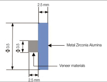
Fig. 2 Specimens for shear bond strength test.

Fig. 3 Schematic diagram of fracture strength test.
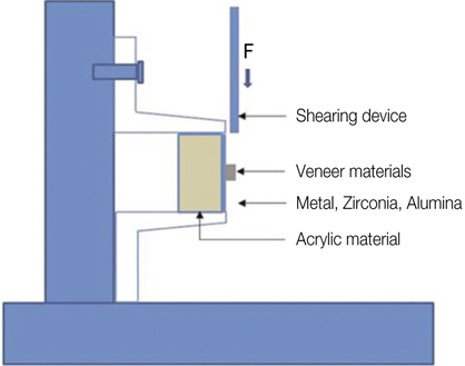
Fig. 4 Schematic diagram of shear bond strength test.
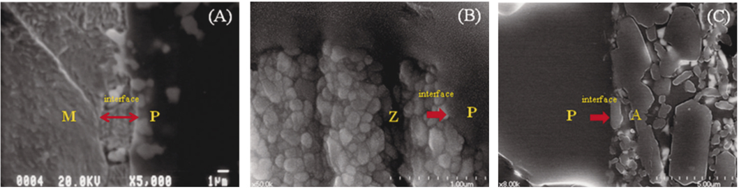
Fig. 5 Cross-sectional SEM image of the interface between porcelain and PFM system (A), zirconia system (B), glass infiltrated alumina system (C). (note : M = metal, P = Porcelain, Z = Zirconia and A = Alumina).
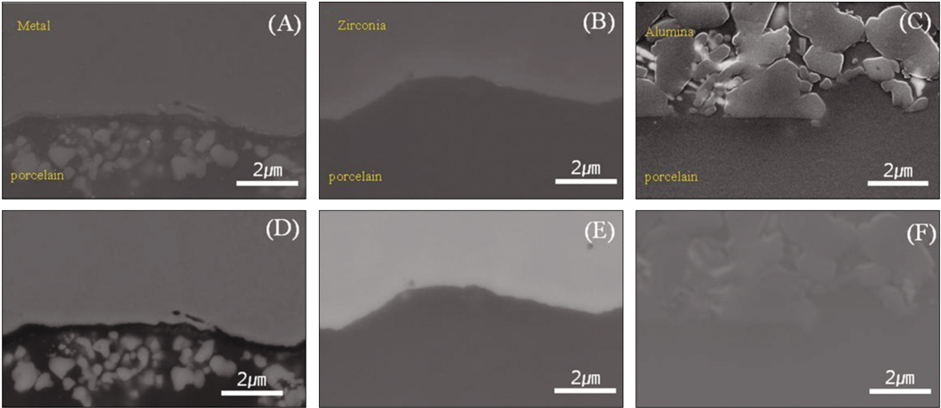
Fig. 6 Cross-sectional SEM image of the interface between porcelain (A) metal, (B) zirconia (C) alumina and back scattered electron image (D), (E), (F). (×50,000).
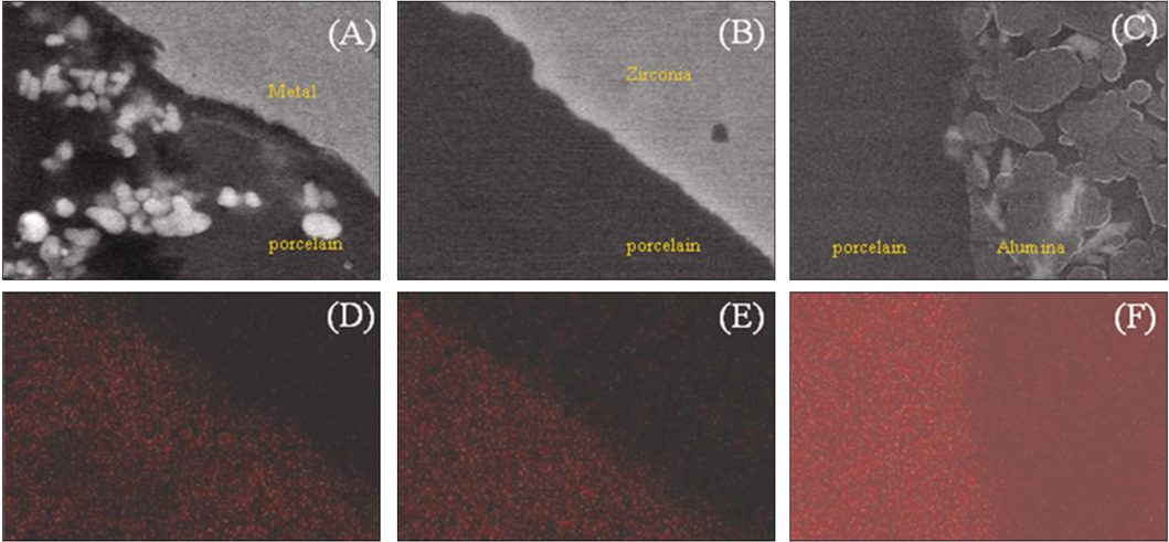
Fig. 7 Cross-sectional SEM image of the interface between porcelain and PFM system (A), zirconia system (B), glass infiltrated alumina system (C) and their EDX mapping image (D, E, F).
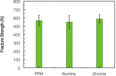
Fig. 8 Fracture strength of experimental groups.

Fig. 9 Fracture pattern of zirconia system (A), glass infiltrated alumina system (B) and PFM system (C).
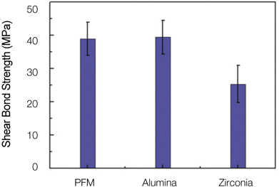
Fig. 10 Shear Bond Strength of experimental groups.
Reference
-
1. Raigrodski AJ. Contemporary materials and technologies for all-ceramic fixed partial dentures: a review of the literature. J Prosthet Dent. 2004. 92:557–562.
Article2. Sproull RC. Color matching in dentistry. Part I. The three-dimensional nature of color. J Prosthet Dent. 1973. 29:416–426.
Article3. Dickinson AJ, Moore BK, Harris RK, Dykema RW. A comparative study of the strength of aluminous porcelain and all-ceramic crowns. J Prosthet Dent. 1989. 61:297–304.
Article4. Levy H, Daniel X. Working with the In-Ceram porcelain system. Prothès Dent. 1990. 44-45:1–11.5. Ludwig K. Investigation of fracture resistance of full porcelain crowns. Dent Labor (Munch). 1991. 39(5):647–651.6. Miller A, Long J, Miller B, Cole J. Comparison of the fracture strengths of ceramometal crowns versus several all-ceramic crowns. J Prosthet Dent. 1992. 68:38–41.
Article7. Rosenstiel SF, Porter SS. Apparent fracture toughness of all-ceramic crown systems. J Prosthet Dent. 1989. 62:529–532.
Article8. Seghi RR, Daher T, Caputo A. Relative flexural strength of dental restorative ceramics. Dent Mater. 1990. 6:181–184.
Article9. Raigrodski AJ, Chiche GJ, Potiket N, Hochstedler JL, Mohamed SE, Billiot S, Mercante DE. The efficacy of posterior three-unit zirconium-oxide-based ceramic fixed partial dental prostheses: a prospective clinical pilot study. J Prosthet Dent. 2006. 96:237–244.
Article10. Sadoun M. All ceramic bridges with the slip casting technique. Presented at the 7th International Symposium on Ceramics. 1988. Paris.11. Al-Makramani BM, Razak AA, Abu-Hassan MI. Biaxial flexural strength of Turkom-Cera core compared to two other all-ceramic systems. J Appl Oral Sci. 2010. 18:607–612.
Article12. AL-Makramani BM, Razak AA, Abu-Hassan MI. Comparison of the load at fracture of Turkom-Cera to Procera AllCeram and In-Ceram all-ceramic restorations. J Prosthodont. 2009. 18:484–488.
Article13. ISO 6872 Dental Ceramic. 2008.14. Anusavice KJ, Carroll JE. Effect of incompatibility stress on the fit of metal-ceramic crowns. J Dent Res. 1987. 66:1341–1345.
Article15. Warpeha WS Jr, Goodkind RJ. Design and technique variables affecting fracture resistance of metal-ceramic restorations. J Prosthet Dent. 1976. 35:291–298.
Article16. Lee IS, Kim JM, Dong JK. Fracture strength of zirconia ceramic crowns according to tooth position. J Korean Acad Prosthodont. 2010. 48:94–100.
Article17. Park HS. A comparative study on the fracture strength of two different CAD/CAM zirconia ceramic system. 2011. In Korea: Seoul National University, College of Dentistry;PhD Thesis.18. Jeong HC. Fracture strength of zirconia monolithic crowns. J Korean Acad Prosthodont. 2006. 44:157–164.19. Hwang JW, Yang JH, Lee SH, Chung HY. A study on fracture strength of conventional and copy-milled In-Ceram crowns. J Korean Acad Prosthodont. 1997. 35:417–430.20. Lee MH, Jeon YC. A study on fracture strength and color by the design if metal coping in ceramo metal crown. J Korean Acad Prosthodont. 1992. 30:103–124.21. Dündar M, Ozcan M, Gökçe B, Cömlekoğlu E, Leite F, Valandro LF. Comparison of two bond strength testing methodologies for bilayered all-ceramics. Dent Mater. 2007. 23:630–636.
Article22. Aboushelib MN, de Jager N, Kleverlaan CJ, Feilzer AJ. Microtensile bond strength of different components of core veneered all-ceramic restorations. Dent Mater. 2005. 21:984–991.
Article23. Aboushelib MN, Kleverlaan CJ, Feilzer AJ. Microtensile bond strength of different components of core veneered all-ceramic restorations. Part II: Zirconia veneering ceramics. Dent Mater. 2006. 22:857–863.
Article24. Aboushelib MN, Kleverlaan CJ, Feilzer AJ. Microtensile bond strength of different components of core veneered all-ceramic restorations. Part 3: double veneer technique. J Prosthodont. 2008. 17:9–13.
Article25. Vickery RC, Badinelli LA. Nature of attachment forces in porcelain-gold systems. J Dent Res. 1968. 47:683–689.
Article26. Knap FJ, Ryge G. Study of bond strength of dental porcelain fused to metal. J Dent Res. 1966. 45:1047–1051.
Article27. Choi BK, Han JS, Yang JH, Lee JB, Kim SH. Shear bond strength of veneering porcelain to zirconia and metal cores. J Adv Prosthodont. 2009. 1:129–135.
Article
- Full Text Links
-
- Actions
-
Cited
- CITED
-
- Close
- Share
-
- Similar articles
-
- COMPARISON OF COLOR AND OPACITY OF COPY-MILLED IN-CREAM ALUMINA CORE AND SPINELL CORE
- MARGINAL FIT OF GLASS INFILTRATED ALUMINA CORE FABRICATED FROM ALUMINA TAPES
- THE PHYSCIAL PORPERTIES OFY Y2O3-CONTAINING GLASS INFILTRATED ALUMINA CORE MADE BY PRESSURELESS POWDER PACKING METHOD
- Marginal fit related to margin types of glass infiltrated alumina core fabricated from aqueous-based alumina tape
- EFFECT OF CeCO2 ADDITION IN GLASS COMPOSITION ON THE STRENGTH OF ALUMINA-GLASS COMPOSITES
