Accuracy assessment of implant placement using a stereolithographic surgical guide made with digital scan
- Affiliations
-
- 1Department of Prosthodontics, Wonju College of Medicine, Yonsei University, Wonju, Republic of Korea. smj3@yonsei.ac.kr
- 2Department of Oral and Maxillofacial Surgery, Wonju College of Medicine, Yonsei University, Wonju, Republic of Korea.
- 3Dio research institute, Pusan, Republic of Korea.
- KMID: 2195393
- DOI: http://doi.org/10.4047/jkap.2015.53.2.111
Abstract
- PURPOSE
The objective of this study was to evaluate the accuracy of a stereolithographic surgical guide that was made with information from intraoral digital impressions and cone beam CT (CBCT).
MATERIALS AND METHODS
Six sets of resin maxilla and mandible models with missing teeth were used in this study. Intraoral digital impressions were made. The virtual models provided by these intraoral digital impressions and by the CBCT scan images of the resin models were used to create a surgical guide. Implant surgery was performed on the resin models using the surgical guide. After implant placement, the models were subjected to another CBCT scan to compare the planned and actual implant positions. Deviations in position, depth and axis between the planned and actual positions were measured for each implant.
RESULTS
The mean deviation of the insertion point and angulation were 0.28 mm and 0.26degrees, apex point were 0.11 mm and 0.14 mm respectively. The implants were situated at a mean of 0.44 mm coronal to the planned vertical position.
CONCLUSION
This study demonstrates that stereolithographic surgical guides created without the use of impressions and stone models show promising accuracy in implant placement.
Figure
-

Fig. 1. Resin maxilla and mandible model with artificial silicone gums.
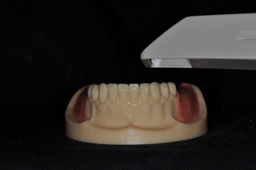
Fig. 2. Intraoral scanning of the resin model.
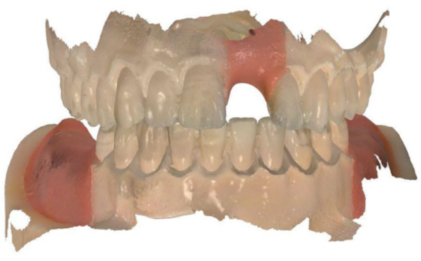
Fig. 3. Scanned model.
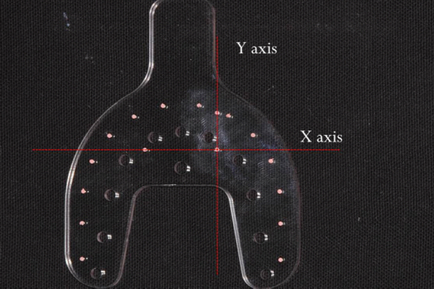
Fig. 4. Plastic plate for determination of a reference plane.

Fig. 5. Plastic plate attached to the resin model.
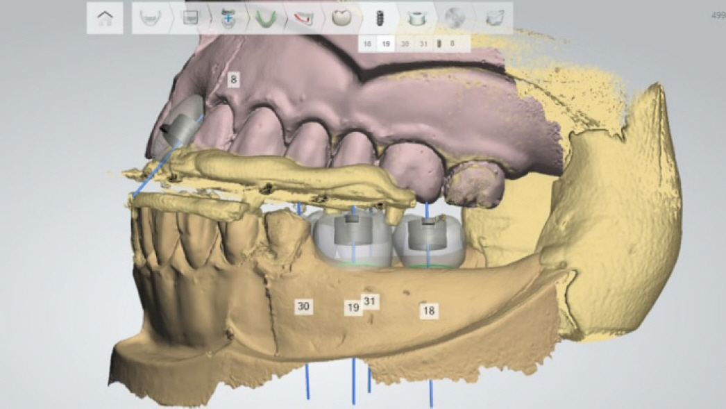
Fig. 6. The merged image of the CBCT scan and the intraoral scan.
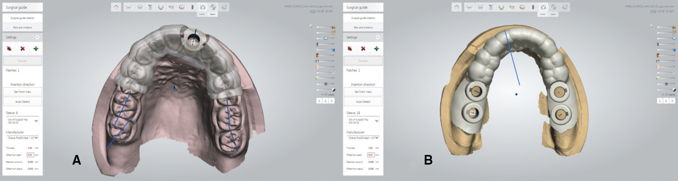
Fig. 7. Virtual surgical guides for the maxilla (A) and mandible (B).
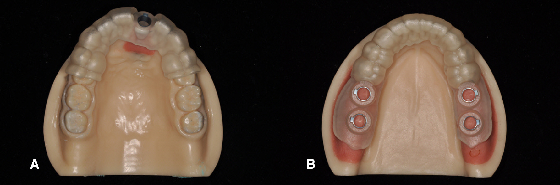
Fig. 8. Surgical guides for the maxilla (A) and mandible (B).
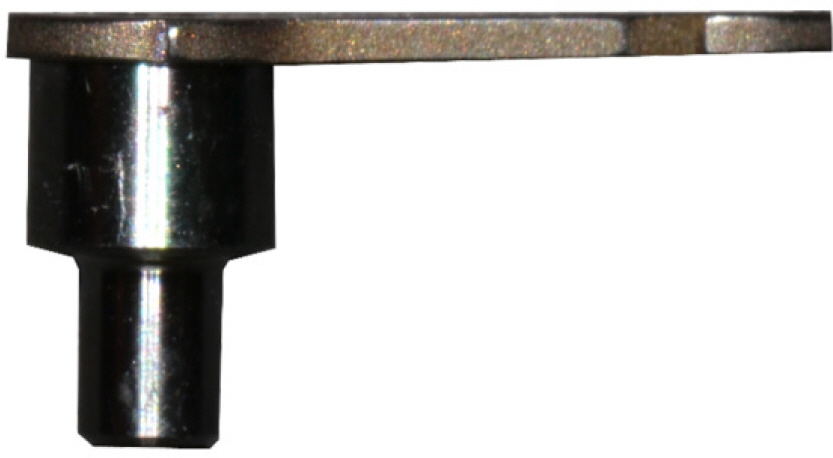
Fig. 9. Guide tube of 9 mm in length.
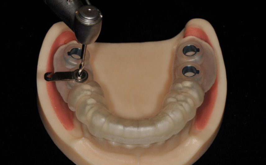
Fig. 10. Drilling through the guide tube with a 2 mm drill.
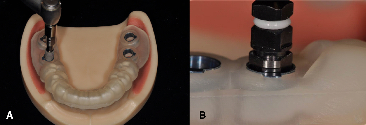
Fig. 11. Surgical guides: Implant placement using the guide (A) and impant connector position at buccal view (B).
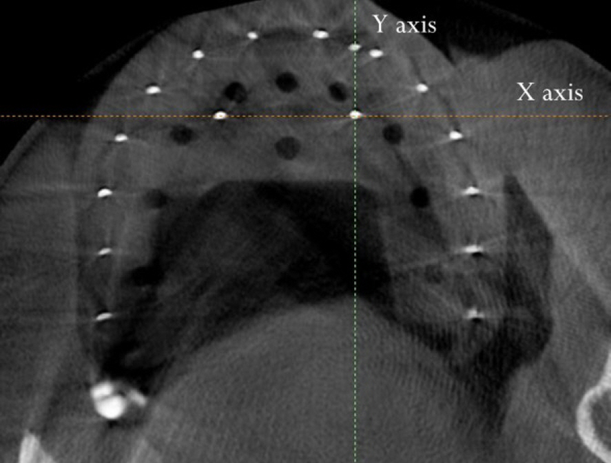
Fig. 12. The X- and Y-axis on the CBCT image.
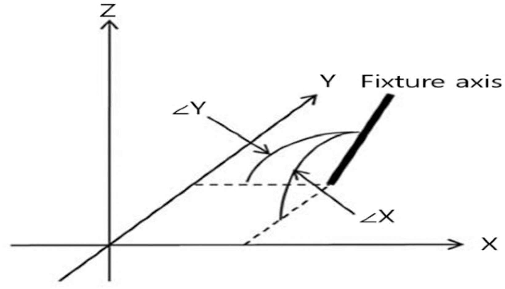
Fig. 13. These illustrations show the procedure used to determine the position and angle of the virtual implant. The insertion point P (X, Y) and apex point P' (X', Y') is determined by the crossing point between the axis of the virtual implant and the XOY-plane. ∠X (Xθ) and ∠Y (Yθ) are defined as the angles from the X- and Y-axes, respectively.
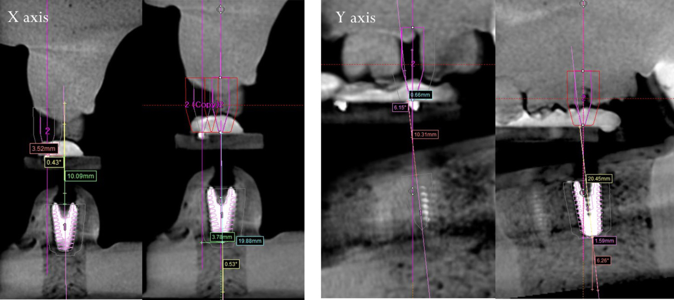
Fig. 14. Position and angulation of the implant on the CBCT image.
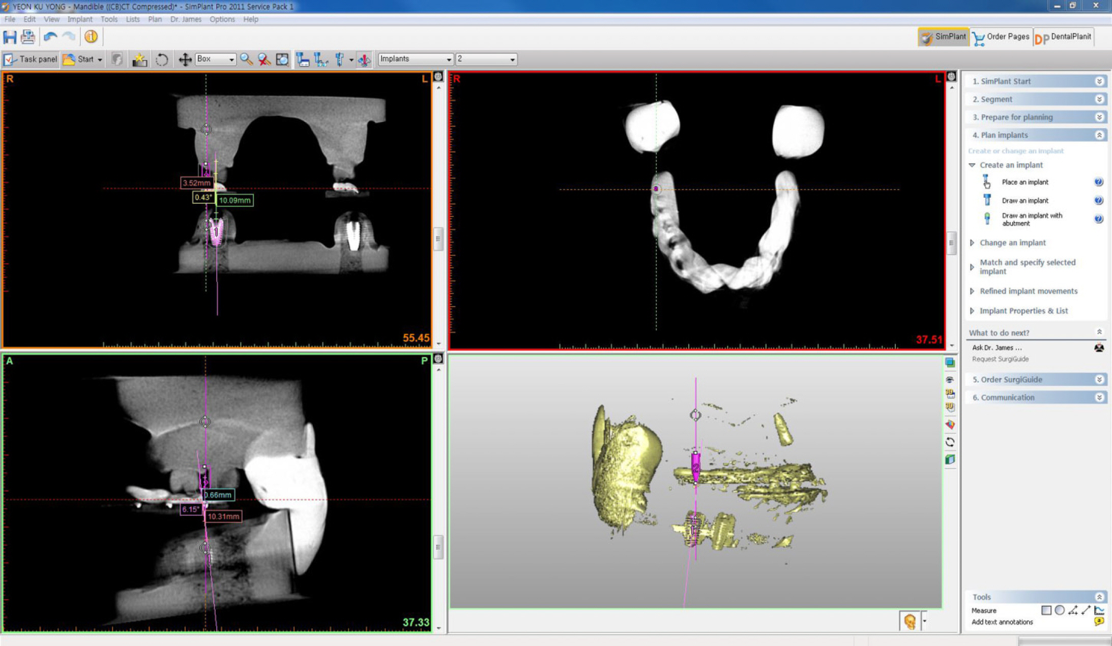
Fig. 15. Position and angulation of the implant on the Simplant software.
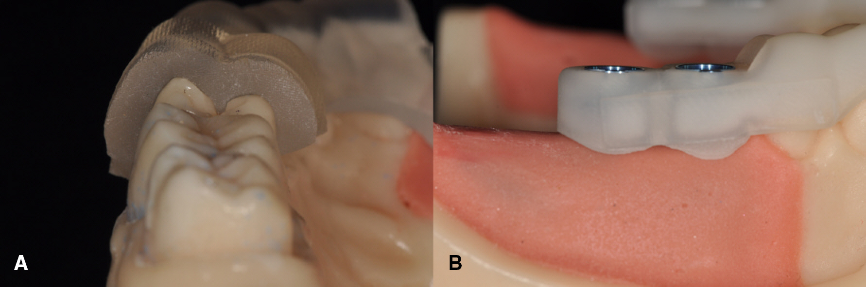
Fig. 16. Surgical guides on the resin maxilla (A) and mandible models (B).
Cited by 1 articles
-
Accuracy of the CT guided implant template by using an intraoral scanner according to the edentulous distance
Byeong-Gil Kang, Hee-Jung Kim, Chae-Heon Chung
J Korean Acad Prosthodont. 2017;55(1):1-8. doi: 10.4047/jkap.2017.55.1.1.
Reference
-
1. Jeong SM, Choi BH, Xuan F, Kim HR. Flapless implant surgery using a mini-incision. Clin Implant Dent Relat Res. 2012; 14:74–9.
Article2. Sclar AG. Guidelines for flapless surgery. J Oral Maxillofac Surg. 2007; 65:20–32.
Article3. Terzioğlu H, Akkaya M, Ozan O. The use of a computerized tomography-based software program with a flapless surgical technique in implant dentistry: a case report. Int J Oral Maxillofac Implants. 2009; 24:137–42.4. Di Giacomo GA, Cury PR, de Araujo NS, Sendyk WR, Sendyk CL. Clinical application of stereolithographic surgical guides for implant placement: preliminary results. J Periodontol. 2005; 76:503–7.5. Ruppin J, Popovic A, Strauss M, Spü ntrup E, Steiner A, Stoll C. Evaluation of the accuracy of three different computer-aided surgery systems in dental implantology: optical tracking vs. stereolithographic splint systems. Clin Oral Implants Res. 2008; 19:709–16.
Article6. Van Assche N, van Steenberghe D, Guerrero ME, Hirsch E, Schutyser F, Quirynen M, Jacobs R. Accuracy of implant placement based on pre-surgical planning of three-dimensional cone-beam images: a pilot study. J Clin Periodontol. 2007; 34:816–21.
Article7. van Steenberghe D, Naert I, Andersson M, Brajnovic I, Van Cleynenbreugel J, Suetens P. A custom template and definitive prosthesis allowing immediate implant loading in the maxilla: a clinical report. Int J Oral Maxillofac Implants. 2002; 17:663–70.8. Kalt G, Gehrke P. Transfer precision of three-dimensional implant planning with CT assisted offline navigation. Int J Comput Dent. 2008; 11:213–25.9. Nickenig HJ, Eitner S. Reliability of implant placement after virtual planning of implant positions using cone beam CT data and surgical (guide) templates. J Craniomaxillofac Surg. 2007; 35:207–11.
Article10. Hajeer MY, Millett DT, Ayoub AF, Siebert JP. Applications of 3D imaging in orthodontics: part II. J Orthod. 2004; 31:154–62.11. Flü gge TV, Nelson K, Schmelzeisen R, Metzger MC. Three-dimensional plotting and printing of an implant drilling guide: simplifying guided implant surgery. J Oral Maxillofac Surg. 2013; 71:1340–6.12. Lee CY, Ganz SD, Wong N, Suzuki JB. Use of cone beam computed tomography and a laser intraoral scanner in virtual dental implant surgery: part 1. Implant Dent. 2012; 21:265–71.13. Stapleton BM, Lin WS, Ntounis A, Harris BT, Morton D. Application of digital diagnostic impression, virtual planning, and computer-guided implant surgery for a CAD/CAM-fabricated, implant-supported fixed dental prosthesis: a clinical report. J Prosthet Dent. 2014; 112:402–8.
Article14. Torassian G, Kau CH, English JD, Powers J, Bussa HI, Marie Salas-Lopez A, Corbett JA. Digital models vs plaster models using alginate and alginate substitute materials. Angle Orthod. 2010; 80:474–81.
Article15. Akyalcin S, Cozad BE, English JD, Colville CD, Laman S. Diagnostic accuracy of impression-free digital models. Am J Orthod Dentofacial Orthop. 2013; 144:916–22.
Article16. Horwitz J, Zuabi O, Machtei E. Radiographic changes around immediately restored dental implants in periodontally susceptible patients: 1-year results. Int J Oral Maxillofac Implants. 2008; 23:531–8.17. Jung RE, Schneider D, Ganeles J, Wismeijer D, Zwahlen M, Hä mmerle CH, Tahmaseb A. Computer technology applications in surgical implant dentistry: a systematic review. Int J Oral Maxillofac Implants. 2009; 24:92–109.
Article
- Full Text Links
-
- Actions
-
Cited
- CITED
-
- Close
- Share
-
- Similar articles
-
- The Accuracy of Implant Placement According to the Height of the Surgical Guide Hole
- Comparison of accuracy between free-hand and surgical guide implant placement among experienced and non-experienced dental implant practitioners: an in vitro study
- Accuracy of digital surgical guides for dental implants
- The use of surgical guide stent for implant placement
- Immediate loading of mandibular single implant by using surgical guide and modeless digital prosthesis: a case report
