The use of surgical guide stent for implant placement
- Affiliations
-
- 1Department of Dentistry, School of Medicine Inha University, Incheon, Republic of Korea. onsdo@inha.ac.kr
- KMID: 2321801
- DOI: http://doi.org/10.4047/jkap.2014.52.4.366
Abstract
- Surgical guide not only provide diagnosis and treatment plan, but even location and direction of implantation. Surgical guide could be divided into non-limited design, partially limited design, and completely limited design. Partially limited design is easily manufactured and inexpensive but less accuracy, compared to completely limited design. From this approach, partially limited design may be particularly effective in patients who present with a single missing tooth or partially edentulous teeth. Completely limited design is anatomically accuracy, esthetical and functional, optimized treatment for prosthetic and biomechanical perspective, and also minimizes discomfort for post-treatment. The purpose of this study is to review previous studies of various surgical guides and applying in clinic.
Figure
-

Fig. 1. Case of #22 implant placement using nonlimiting design surgical template. (A) Missing tooth of #22 was seen in panoramic view of pre-surgery. (B) Surgical template made by clear vacuum formed matrix on the cast. (C) Try in the template to the patient's mouth. (D) Implant was placed properly in panoramic view of post-surgery.
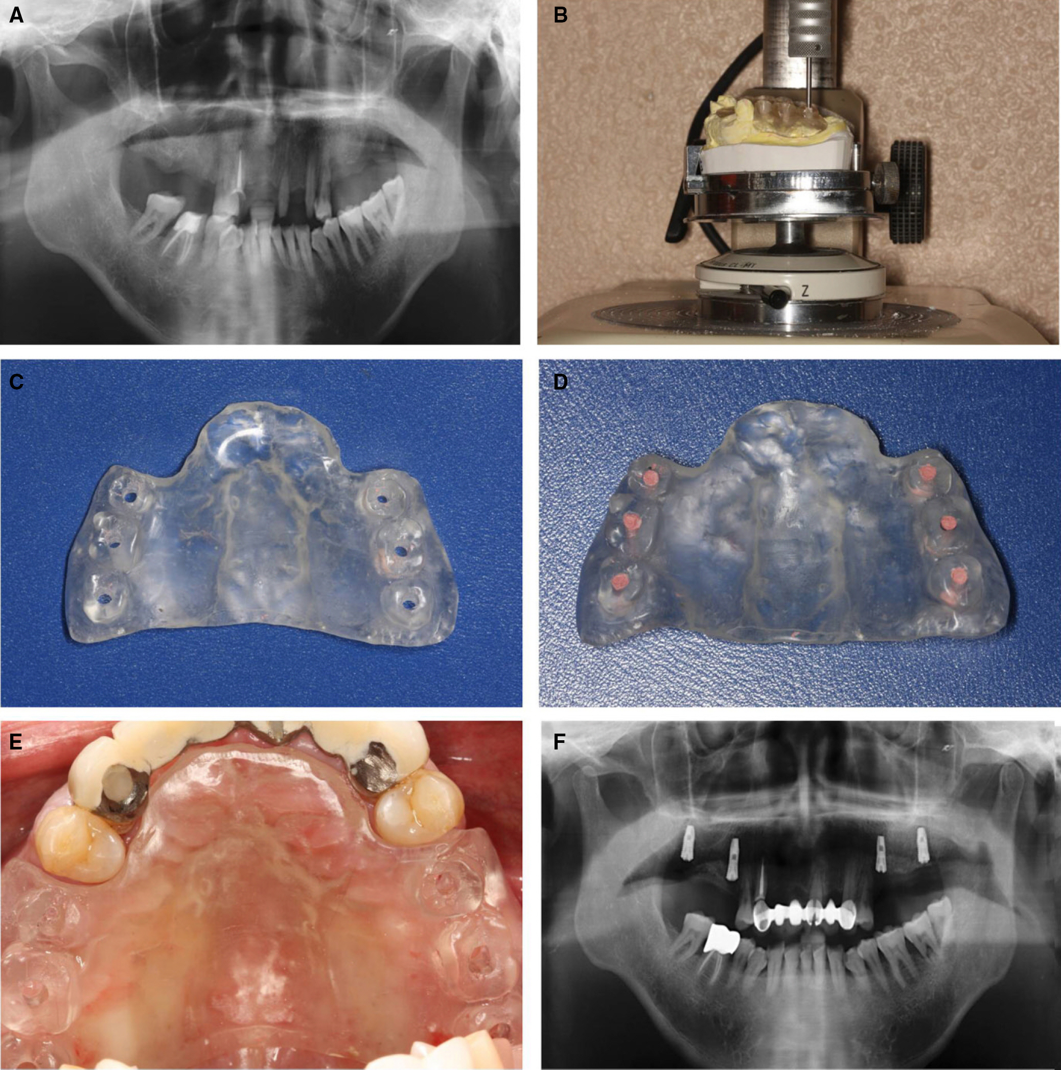
Fig. 2. Case of implant placement on both side of maxillary molar by using resin stent. (A) The patient has maxillary partial edentulous molar area on both side, therefore treatment plan was set by four implants and fixed prosthesis. (B) Resin template was fabricated with cast and pouring resin. Put the template on the surveyor and drilling in the appropriate implant path with long shank round bur. (C) The implant drill hole was formed for appropriate path and direction. (D) Holes were filled with Gutta percha for CT taking. Appropriate direction and position for anatomical condition was checked by cone beam CT. (E) The stent was used as guide for drilling position and direction in implant surgery. (F) Four implants were placed in good direction and position on both side of maxillary molar area.

Fig. 3. Case of implant placement in #22 by using thermo-plastic polymer guide (Dentium Thermo-plastic stent;Dentium Korea). (A) Left maxillary lateral incisor was missing and planned to treat with implant. For the stent making, cast model was drilled in appropriate position and direction and inserted guide pin. (B) Thermo-Plastic stent form before shaping. (C) Put the stent into hot water for shaping. (D) The polymer guide was made appropriately and applied onto plaster cast. (E) Position the polymer guide to the surgical site and drilling for implantation. (F) By using simply made surgical guide, the implant fixture was placed in well direction and position.
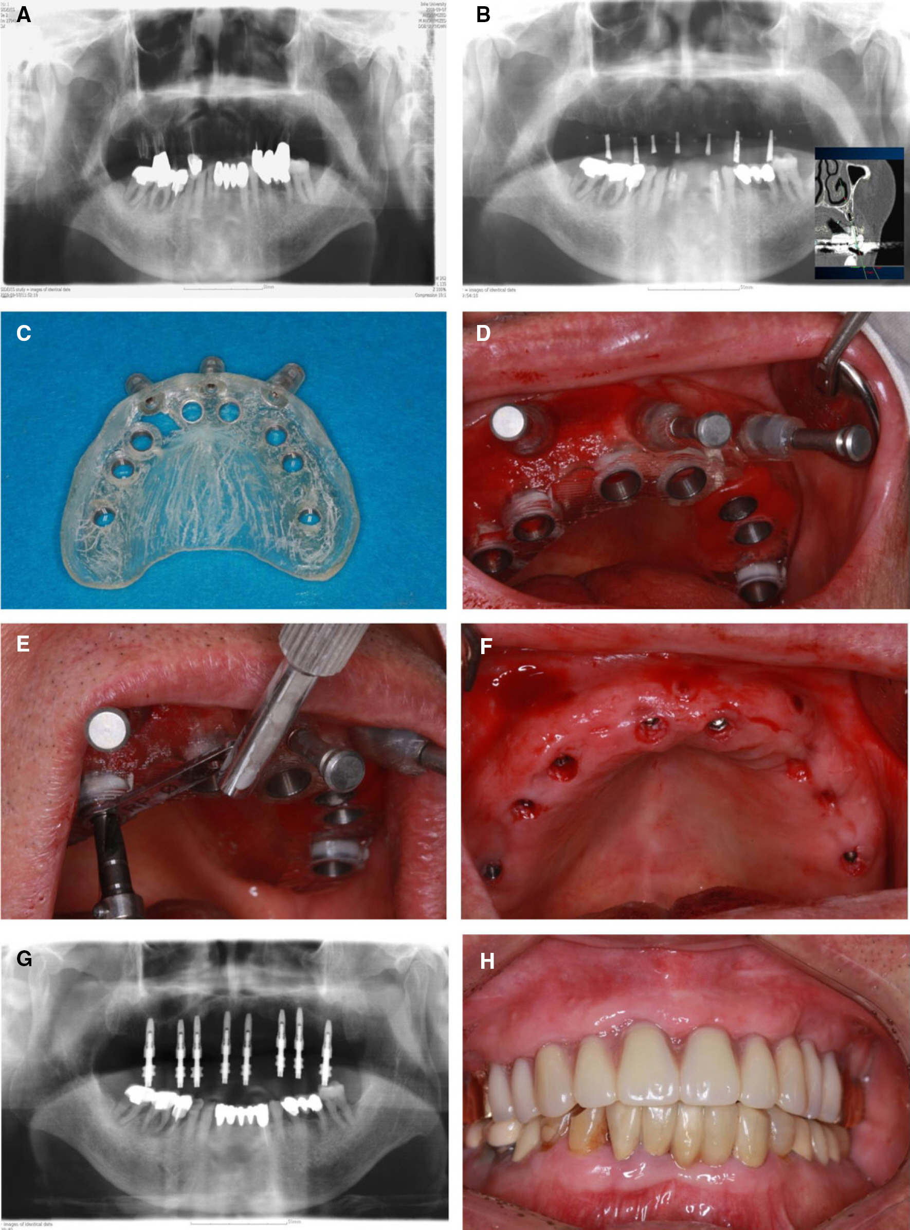
Fig. 4. Case of full maxillary rehabilitation by implants with Nobel Guide Surgical Kit (Nobelbiocare, USA). (A) Because of severe chronic periodontitis, all remaining maxillary teeth were planned to extract. Rehabilitation of maxillary defect with eight implants and fixed prosthesis was planned. (B) Conventional resin template was made. The patient was delivered template, and taken panoramic radiography and cone beam CT for the position and direction of implant placement. CT images was loaded by NobelGuideTM software and reconstructed on 3D. The diameter of fixture and position of implant placement was decided by using remain bone and noninvasion of anatomical structure. (C) NobelGuide stent which is set the position and direction was made by analysis results. (D) Locate the surgical stent using surgical index. Stable the stent with anchor pin. (E) Tailored drill guides are available for each drill diameter to lead the guided drills through the surgical template. (F) Eight implants were placed in maxilla by using NobelGuide stent with flapless surgery. (G) Implant coping was inserted for prosthesis making, taken panoramic radiograph and impression. (H) Maxilla was rehabilitated by implant supported fixed prosthesis.
Cited by 1 articles
-
Implant overdenture of mandible with severe unilateral atrophy: Report of two cases
So-Yeun Kim, Eun-Young Kwon, Kyoung-Hwa Jung, Hye-Mi Jeon, Young-Jae Baek, Mi-Jung Yun, Jung-Bo Huh
J Korean Acad Prosthodont. 2019;57(3):271-279. doi: 10.4047/jkap.2019.57.3.271.
Reference
-
1. Pattanaik S, Pattanaik BK. Fabrication of a surgical guide with help of a milling machine by ridge mapping method. J Indian Prosthodont Soc. 2013; 13:61–5.
Article2. Carl CE. Misch. Diagnostic model and surgical guide.1st ed., Dent Implant Prosthetics. Elsevier Science Health;USA: 2005. p. 54–68. p. 142–55.3. Park HW, Park SJ, Kim MS, Park HJ. Computerized planning of dental implant placement using geometric processing of 3D models. J Comput Des Eng. 2011. 955–9.4. D'Souza KM, Aras MA. Types of implant surgical guides in dentistry: a review. J Oral Implantol. 2012; 38:643–52.5. Blustein R, Jackson R, Rotskoff K, Coy RE, Godar D. Use of splint material in the placement of implants. Int J Oral Maxillofac Implants. 1986; 1:47–9.6. Engelman MJ, Sorensen JA, Moy P. Optimum placement of osseointegrated implants. J Prosthet Dent. 1988; 59:467–73.
Article7. Stumpel LJ 3rd. Cast-based guided implant placement: a novel technique. J Prosthet Dent. 2008; 100:61–9.
Article8. Lee DH. New method to fabricate correct surgical stent in implant surgery. J Korean Dent Assoc. 1993; 31:183–9.9. Adrian ED, Ivanhoe JR, Krantz WA. Trajectory surgical guide stent for implant placement. J Prosthet Dent. 1992; 67:687–91.
Article10. Tarlow JL. Fabrication of an implant surgical stent for the edentulous mandible. J Prosthet Dent. 1992; 67:217–8.
Article11. Espinosa Marino J, Alvarez Arenal A, Pardo Ceballos A, Fernandez Vazquez JP, Ibaseta Diaz G. Fabrication of an implant radiologic-surgical stent for the partially edentulous patient. Quintessence Int. 1995; 26:111–4.12. Stellino G, Morgano SM, Imbelloni A. A dual-purpose, implant stent made from a provisional fixed partial denture. J Prosthet Dent. 1995; 74:212–4.
Article13. Pesun IJ, Gardner FM. Fabrication of a guide for radiographic evaluation and surgical placement of implants. J Prosthet Dent. 1995; 73:548–52.
Article14. Takeshita F, Tokoshima T, Suetsugu T. A stent for presurgical evaluation of implant placement. J Prosthet Dent. 1997; 77:36–8.
Article15. Sicilia A, Noguerol B, Cobo J, Zabalegui I. Profile surgical template: a systematic approach to precise implant placement. A technical note. Int J Oral Maxillofac Implants. 1998; 13:109–14.16. Minoretti R, Merz BR, Triaca A. Predetermined implant positioning by means of a novel guide template technique. Clin Oral Implants Res. 2000; 11:266–72.
Article17. Becker CM, Kaiser DA. Surgical guide for dental implant placement. J Prosthet Dent. 2000; 83:248–51.
Article18. Almog DM, Torrado E, Meitner SW. Fabrication of imaging and surgical guides for dental implants. J Prosthet Dent. 2001; 85:504–8.
Article19. Lee JH, Kim SM, Paeng JY, Kim MJ. Implant surgery based on computer simulation surgical stent and the assessment with the image fusion technique. J Korean Assoc Oral Maxillofac Surg. 2010; 36:402–7.
Article20. Huynh-Ba G, Alexander P, Vargas A, Vierra M, Oates TW. A radiographic template for a two-implant mandibular overdenture using the patient's existing denture. J Prosthet Dent. 2013; 109:53–6.21. Moslehifard E, Nokar S. Designing a custom made gauge device for application in the access hole correction in the dental implant surgical guide. J Indian Prosthodont Soc. 2012; 12:123–9.
Article22. Kim KG, Ji HK, Yang EK, Bae YS. 3D Image guided implant surgery system by fusion of stent and image guided-surgery. J Comput Sci Eng. 2008; 35:268–71.23. Wanschitz F, Birkfellner W, Watzinger F, Schopper C, Patruta S, Kainberger F, Figl M, Kettenbach J, Bergmann H, Ewers R. Evaluation of accuracy of computer-aided intraoperative positioning of endosseous oral implants in the edentulous mandible. Clin Oral Implants Res. 2002; 13:59–64.
Article24. Van Steenberghe D, Malevez C, Van Cleynenbreugel J, Bou Serhal C, Dhoore E, Schutyser F, Suetens P, Jacobs R. Accuracy of drilling guides for transfer from three-dimensional CT-based planning to placement of zygoma implants in human cadavers. Clin Oral Implants Res. 2003; 14:131–6.
Article25. Fortin T, Coudert JL, Champleboux G, Sautot P, Lavalle′e S. Computer-assisted dental implant surgery using computed tomography. J Image Guid Surg. 1995; 1:53–8.
Article26. Noharet R, Pettersson A, Bourgeois D. Accuracy of implant placement in the posterior maxilla as related to 2 types of surgical guides: a pilot study in the human cadaver. J Prosthet Dent. 2014; 112:526–32.27. Kwon CR, Choi BH, Jeong SM, Joo SD. Evaluation of the accuracy of two different surgical guides in dental implantology: stereolithography fabricated vs. positioning device fabricated surgical guides. J Korean Acad Prosthodont. 2012; 50:271–8.
Article28. Stellino G, Morgano SM, Imbelloni A. A dual-purpose, implant stent made from a provisional fixed partial denture. J Prosthet Dent. 1995; 74:212–4.
Article
- Full Text Links
-
- Actions
-
Cited
- CITED
-
- Close
- Share
-
- Similar articles
-
- The Accuracy of Implant Placement According to the Height of the Surgical Guide Hole
- Clinical Precautions for Implant Placement using Computer-guided Implant Surgical Guide: A Systematic Review
- Implant-supported milled bar overdenture with two implant surgical guides
- An assessment of accuracy of half-guided implant surgery using implant surgical guide: A case report
- A case report of a surgical guide fabricated via intraoral scanning-based implant planning and wax-based rapid prototyping
