Periorbital changes with aging
- Affiliations
-
- 1Department of Ophthalmology, Seoul Metropolitan Government Seoul National University Boramae Medical Center, Seoul National University College of Medicine, Seoul, Korea. hokyung214@gmail.com
- KMID: 2193476
- DOI: http://doi.org/10.5124/jkma.2013.56.11.1012
Abstract
- As we face a rapidly aging population in the Republic of Korea, the number of patients with the need to prevent or alleviate periorbital changes due to aging will grow. The periorbital changes that occur with aging comprise a dynamic process involving the aging of facial tissue and bony structures. Epidermal thinning and decreases in collagen cause the skin to lose its elasticity. Loss of fat, coupled with gravity and muscle pull, leads to wrinkling and the formation of dynamic lines. The aging process has also been shown to affect the facial bones. Multiple studies suggest that aging of the orbit and midface bones occurs primarily due to contraction and morphologic changes. This loss of bony volume and projection may contribute to an aged appearance. The effort to understand each patient's individual involutional changes, which differ by age and gender, is mandatory. Identifying the patient's personal needs and selecting the appropriate treatment accordingly is crucial for achieving the best outcome both for the clinician and the patient.
Keyword
MeSH Terms
Figure
-
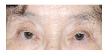
Figure 1 Photograph of a 74-year-old woman with high placement of multiple creases and hollow superior sulcus.

Figure 2 Photograph showing upper lid ptosis (asterisk) with elevated eyebrow and forehead creases (arrow).
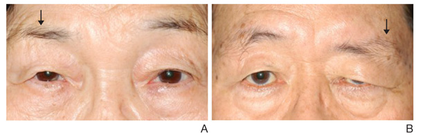
Figure 3 Photographs showing asymmetric eyebrow (arrow) in patients with history of facial nerve palsy. (A) Photograph of right brow ptosis with history of right facial nerve palsy. (B) Photograph of left brow ptosis with history of left facial nerve palsy.
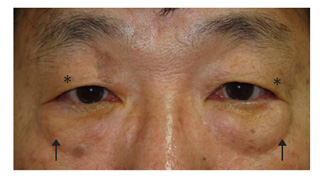
Figure 4 Photographs of a 57-year-old man showing both upper lid skin drooping (dermatochalasis, asterisk) and prolapsed fat on both lower eyelids (festoon, arrow).
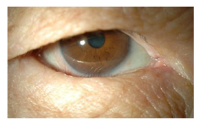
Figure 5 Photograph of entropion in a 66-year-old woman showing inversion of the lower eyelid with cilia and eyelid skin touching the cornea.
Reference
-
1. Lott P, Caldiera AM, Lucas A, Grigalek G. Envejecimiento facial. Papel de la orbita senil. Cir Plast Ibereo-Latinoame. 1996; 22:21–30.2. Loeb R. Anatomical considerations. In : Loeb R, editor. Aesthetic surgery of the eyelids. New York: Springer-Verlag;1989. p. 1–12.3. Matsuo K, Kondoh S, Kitazawa T, Ishigaki Y, Kikuchi N. Pathogenesis and surgical correction of dynamic lower scleral show as a sign of disinsertion of the levator aponeurosis from the tarsus. Br J Plast Surg. 2005; 58:668–675.
Article4. Sultana R, Matsuo K, Yuzuriha S, Kushima H. Disinsertion of the levator aponeurosis from the tarsus in growing children. Plast Reconstr Surg. 2000; 106:563–570.
Article5. Har-Shai Y, Hirshowitz B. Extended upper blepharoplasty for lateral hooding of the upper eyelid using a scalpel-shaped excision: a 13-year experience. Plast Reconstr Surg. 2004; 113:1028–1035.
Article6. Shore JW, McCord CD Jr. Anatomic changes in involutional blepharoptosis. Am J Ophthalmol. 1984; 98:21–27.
Article7. Park CY, Jeon SL, Woo KI, Chang HR. The frequency and aspects of ptosis in Korean old age. J Korean Ophthalmol Soc. 2007; 48:205–210.8. Sanke RF. Relationship of senile ptosis to age. Ann Ophthalmol. 1984; 16:928–931.9. Goldberg B, Rabinovitch M. Connective tissue. In : Weiss L, Greep RO, editors. Histology. 4th ed. New York: McGraw-Hill;1977. p. 145–178.10. Bailey AJ, Duance VC. Collagen in acquired connective tissue diseases: an active or passive role? Eur J Clin Invest. 1980; 10:1–3.
Article11. Matros E, Garcia JA, Yaremchuk MJ. Changes in eyebrow position and shape with aging. Plast Reconstr Surg. 2009; 124:1296–1301.
Article12. Seo HR, Ahn HB. Morphological changes of the eyelid according to age. J Korean Ophthalmol Soc. 2009; 50:1461–1467.
Article13. American Academy of Ophthalmology. Anatomy. American Academy of Ophthalmology. Orbit, eyelids, and lacrimal system, 2006-2007. San Francisco: American Academy of Ophthalmology;2006. p. 135–147.14. Cantisano-Zilkha M. Patient home care regimens. Ophthalmol Clin North Am. 2005; 18:325–327.
Article15. Mendelson BC, Hartley W, Scott M, McNab A, Granzow JW. Age-related changes of the orbit and midcheek and the implications for facial rejuvenation. Aesthetic Plast Surg. 2007; 31:419–423.
Article16. Benedetto AV. The environment and skin aging. Clin Dermatol 1. Clin Dermatol. 1998; 16:129–139.
- Full Text Links
-
- Actions
-
Cited
- CITED
-
- Close
- Share
-
- Similar articles
-
- A Case of Conjunctival and Periorbital Hemorrhage Spontaneously Occurred in Clopidogrel-treated Patient
- Usefulness of Gold Thread Implantation for Crow's Feet
- Two Cases of Periorbital Necrotizing Fasciitis in Immunocompromised Patients
- Can we rejuvenate? Implications of biological aging research
- Periorbital Lipogranuloma after Autologous Fat Injection for Facial Augmentation
