Complete Separation of the Vertebral Body Associated with a Schmorl's Node Accompanying Severe Osteoporosis
- Affiliations
-
- 1Department of Natural Medical Sciences, College of Health Science, Chosun University, Gwangju, Korea.
- 2Department of Neurosurgery, Heori Sarang Hospital, Daejeon, Korea.
- 3Department of Internal Medicine, Soonchunhyang University College of Medicine, Seoul, Korea.
- 4Department of Neurosurgery, College of Medicine, Chosun University, Gwangju, Korea. chosunns@chosun.ac.kr
- KMID: 2191325
- DOI: http://doi.org/10.3340/jkns.2015.58.2.147
Abstract
- A Schmorl's node is defined as a simple endplate intravertebral herniation resulting from trauma or idiopathic causes. Although Schmorl's nodes have been considered clinically insignificant, they might indicate an active symptomatic process or cause serious complications. In this study, we report an interesting case of complete separation of a vertebral body caused by an untreated Schmorl's node accompanying severe osteoporosis. To our knowledge, this is the first clinical report in the published literature to evaluate the complete separation of a vertebral body associated with a Schmorl's node.
Keyword
MeSH Terms
Figure
-
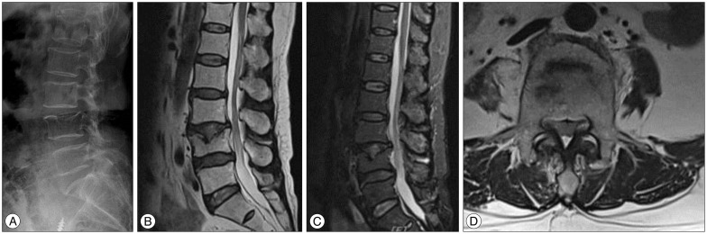
Fig. 1 A symptomatic Schmorl's node arising after a trivial injury in a 67-year-old woman. A : Simple lateral radiograph at the time of initial injury reveals minimal bony change at the middle and posterior endplate of L4. B, C and D : Sagittal T2, fat suppression, and axial T2 magnetic resonance images present a minimal Schmorl's node at L4 and surrounding edematous change.

Fig. 2 A developed Schmorl's node with complete separation of the vertebral body after 18 months. A : Simple lateral radiograph shows separation of the vertebral body at L4. B and C : Follow-up magnetic resonance images reveal a large defect and fragmentation within the vertebral body with concurrent spinal stenosis. D : Computed tomography reveals complete separation of the vertebral body at L4.
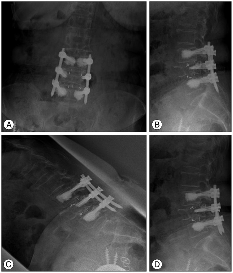
Fig. 3 Simple anteroposterior and lateral (A and B) radiographs and dynamic flexion and extension views (C and D) taken 2 years after bone cement augmented percutaneous posterior lumbar interbody fusion at L3-4 and L4-5.
Reference
-
1. Coventry MB, Ghormley RK, Kernohan JW. The intervertebral disc : its microscopic anatomy and pathology : part II. changes in the intervertebral disc concomitant with age. J Bone Joint Surg Am. 1945; 27:233–247.2. Coventry MB, Ghormley RK, Kernohan JW. The intervertebral disc : its microscopic anatomy and pathology : part III. pathological changes in the intervertebral disc. J Bone Joint Surg Am. 1945; 27:460–474.3. Dimar JR 2nd, Nathan ST, Glassman SD. The spectrum of traumatic Schmorl's nodes : identification and treatment options in 3 patients. Am J Orthop (Belle Mead NJ). 2012; 41:427–431. PMID: 23365812.4. Fahey V, Opeskin K, Silberstein M, Anderson R, Briggs C. The pathogenesis of Schmorl's nodes in relation to acute trauma. An autopsy study. Spine (Phila Pa 1976). 1998; 23:2272–2275. PMID: 9820905.5. Hamanishi C, Kawabata T, Yosii T, Tanaka S. Schmorl's nodes on magnetic resonance imaging. Their incidence and clinical relevance. Spine (Phila Pa 1976). 1994; 19:450–453. PMID: 8178234.6. Hasegawa K, Ogose A, Morita T, Hirata Y. Painful Schmorl's node treated by lumbar interbody fusion. Spinal Cord. 2004; 42:124–128. PMID: 14765146.
Article7. Kim HS, Kim SH, Ju CI, Kim SW, Lee SM, Shin H. The role of bone cement augmentation in the treatment of chronic symptomatic osteoporotic compression fracture. J Korean Neurosurg Soc. 2010; 48:490–495.
Article8. Masala S, Pipitone V, Tomassini M, Massari F, Romagnoli A, Simonetti G. Percutaneous vertebroplasty in painful schmorl nodes. Cardiovasc Intervent Radiol. 2006; 29:97–101. PMID: 16328689.
Article9. Möller A, Maly P, Besjakov J, Hasserius R, Ohlin A, Karlsson MK. A vertebral fracture in childhood is not a risk factor for disc degeneration but for Schmorl's nodes : a mean 40-year observational study. Spine (Phila Pa 1976). 2007; 32:2487–2492. PMID: 18090090.
Article10. Resnick D, Niwayama G. Intravertebral disk herniations : cartilaginous (Schmorl's) nodes. Radiology. 1978; 126:57–65. PMID: 339268.
Article11. Takahashi K, Miyazaki T, Ohnari H, Takino T, Tomita K. Schmorl's nodes and low-back pain. Analysis of magnetic resonance imaging findings in symptomatic and asymptomatic individuals. Eur Spine J. 1995; 4:56–59. PMID: 7749909.12. Takahashi K, Takata K. A large painful Schmorl's node : a case report. J Spinal Disord. 1994; 7:77–81. PMID: 8186593.
- Full Text Links
-
- Actions
-
Cited
- CITED
-
- Close
- Share
-
- Similar articles
-
- Schmorl’s Node Accompanying Multiple Osteonecroses
- Rami Communicans Nerve Block for the Treatment of Symptomatic Schmorl's Nodes: A Case Report
- Acute Schmorl Node in Dorsal Spine: An Unusual Cause of a Sudden Onset of Severe Back Pain in a Young Female
- Papillary Thyroid Carcinoma Metastasis to the Lumbar Spine Masquerading as a Schmorl's Node
- Pregnancy and Lactation-associated Osteoporosis with Vertebral Compression Fracture
