Primary Osteolytic Intraosseous Atypical Meningioma with Soft Tissue and Dural Invasion: Report of a Case and Review of Literatures
- Affiliations
-
- 1Department of Neurosurgery, Dankook University College of Medicine, Cheonan, Korea. lsk999999@gmail.com
- KMID: 2191146
- DOI: http://doi.org/10.3340/jkns.2014.56.6.509
Abstract
- Primary intraosseous meningioma is a rare tumor, and atypical pathologic components both osteolytic lesion and dura and soft tissue invasion is extremely rare. A 65-year-old woman presented with a 5-month history of a soft mass on the right frontal area. MR imaging revealed a 4 cm sized, multilobulated, strongly-enhancing lesion on the right frontal bone, and CT showed a destructive skull lesion. The mass was adhered tightly to the scalp and dura mater, and it extended to some part of the outer and inner dural layers without brain invasion. The extradural mass and soft tissue mass were totally removed simultaneously and we reconstructed the calvarial defect with artificial bone material. The pathological study revealed an atypical meningioma as World Health Organization grade II. Six months after the operation, brain MR imaging showed that not found recurrence in both cranial and spinal lesion. Here, we report a case of primary osteolytic intraosseous atypical meningioma with soft tissue and dural invasion.
Keyword
MeSH Terms
Figure
-
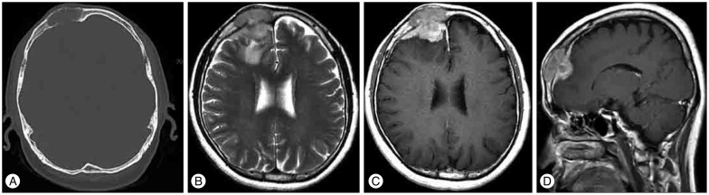
Fig. 1 Initial CT and MR scans. CT scan with bone window (A) demonstrates a right-sided frontal mass expanding the calvaria with cortical destruction. Axial T2-weighted (B) MR images show the frontal intracalvarial mass lesion that was hypointense on T2-weighted images with the surrounding edema. The lesion shows intense and homogeneous enhancement on the postcontrast T1-weighted (C) image. Sagittal postcontrast T1-weighted MR images (D) reveal the intracranial extension and extradural location of the lesion.

Fig. 2 Pathologic findings of the tumor specimens. Histological specimen showing atypical meningioma with freguent mitosis (A) and geographic necrosis (B) (H&E, original magnification ×100).
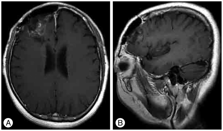
Fig. 3 Follow-up images 6 months after the operation. Postoperative MR image axial postcontrast T1-weighted (A) and sagittal postcontrast T1-weighted (B) MR images show no recurrence of the mass lesion except for postoperative changes.
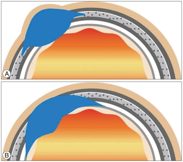
Fig. 4 Comparative illustrations of the tumor of bone origin including intraosseous meningioma (A) and the tumor of meningeal origin including intracranial meningioma (B). Prior illustration (A), which demonstrates a broad calvarial tumor base, shows more expansion of the soft tissue and dural lesion while tumor base of the next illustration (B) located in the meaninges including the dura or arachnoid and it shows more expansion of the brain, bone and a few soft tissue lesions.
Cited by 1 articles
-
Primary Intraosseous Osteolytic Meningioma of the Skull Mimicking Scalp Mass: A Case Report and Review of Literature
Soon Young Kwon, Hyung Shik Shin, Tae Hong Kim, Hyun Jung Kim
Brain Tumor Res Treat. 2015;3(2):151-155. doi: 10.14791/btrt.2015.3.2.151.
Reference
-
1. Arana E, Diaz C, Latorre FF, Menor F, Revert A, Beltrán A, et al. Primary intraosseous meningiomas. Acta Radiol. 1996; 37:937–942. PMID: 8995470.
Article2. Azar-Kia B, Sarwar M, Marc JA, Schechter MM. Intraosseous meningioma. Neuroradiology. 1974; 6:246–253. PMID: 4810615.
Article3. Bassiouni H, Asgari S, Hübschen U, König HJ, Stolke D. Dural involvement in primary extradural meningiomas of the cranial vault. J Neurosurg. 2006; 105:51–59. PMID: 16871880.
Article4. Borggreven PA, de Graaf FH, van der Valk P, Leemans CR. Post-traumatic cutaneous meningioma. J Laryngol Otol. 2004; 118:228–230. PMID: 15068523.
Article5. Changhong L, Naiyin C, Yuehuan G, Lianzhong Z. Primary intraosseous meningiomas of the skull. Clin Radiol. 1997; 52:546–549. PMID: 9240709.
Article6. Crawford TS, Kleinschmidt-DeMasters BK, Lillehei KO. Primary intraosseous meningioma. Case report. J Neurosurg. 1995; 83:912–915. PMID: 7472564.7. Elder JB, Atkinson R, Zee CS, Chen TC. Primary intraosseous meningioma. Neurosurg Focus. 2007; 23:E13. PMID: 17961037.
Article8. Guermazi A, Lafitte F, Miaux Y, Adem C, Bonneville JF, Chiras J. The dural tail sign--beyond meningioma. Clin Radiol. 2005; 60:171–188. PMID: 15664571.
Article9. Hoye SJ, Hoar CS Jr, Murray JE. Extracranial meningioma presenting as a tumor of the neck. Am J Surg. 1960; 100:486–489. PMID: 13716296.
Article10. Jayaraj K, Martinez S, Freeman A, Lyles KW. Intraosseous meningioma-- a mimicry of Paget’s disease. J Bone Miner Res. 2001; 16:1154–1156. PMID: 11393793.11. Kim H, Jung TY, Kim IY, Lee JK. Two cases of primary osteolytic intraosseous meningioma of the skull metastasizing to whole skull and the spine. J Korean Neurosurg Soc. 2012; 51:151–154. PMID: 22639712.
Article12. Lang FF, Macdonald OK, Fuller GN, DeMonte F. Primary extradural meningiomas : a report on nine cases and review of the literature from the era of computerized tomography scanning. J Neurosurg. 2000; 93:940–950. PMID: 11117866.
Article13. Marwah N, Gupta S, Marwah S, Singh S, Kalra R, Arora B. Primary intraosseous meningioma. Indian J Pathol Microbiol. 2008; 51:51–52. PMID: 18417855.
Article14. Mattox A, Hughes B, Oleson J, Reardon D, McLendon R, Adamson C. Treatment recommendations for primary extradural meningiomas. Cancer. 2011; 117:24–38. PMID: 20824719.
Article15. Partington MD, Scheithauer BW, Piepgras DG. Carcinoembryonic antigen production associated with an osteolytic meningioma. Case report. J Neurosurg. 1995; 82:489–492. PMID: 7861230.
Article16. Politi M, Romeike BF, Papanagiotou P, Nabhan A, Struffert T, Feiden W, et al. Intraosseous hemangioma of the skull with dural tail sign : radiologic features with pathologic correlation. AJNR Am J Neuroradiol. 2005; 26:2049–2052. PMID: 16155158.17. Rokni-Yazdi H, Sotoudeh H. Prevalence of "dural tail sign" in patients with different intracranial pathologies. Eur J Radiol. 2006; 60:42–45. PMID: 16675180.
Article18. Shuangshoti S. Primary meningiomas outside the central nervous system. In : Al-Mefty O, editor. Meningiomas. New York, NY: Raven Press;1991. p. 107–128.19. Siegel GJ, Anderson PJ. Extracalvarial meningioma. Case report. J Neurosurg. 1966; 25:83–86. PMID: 5947052.20. Tokgoz N, Oner YA, Kaymaz M, Ucar M, Yilmaz G, Tali TE. Primary intraosseous meningioma : CT and MRI appearance. AJNR Am J Neuroradiol. 2005; 26:2053–2056. PMID: 16155159.
- Full Text Links
-
- Actions
-
Cited
- CITED
-
- Close
- Share
-
- Similar articles
-
- Two Cases of Primary Osteolytic Intraosseous Meningioma of the Skull Metastasizing to Whole Skull and the Spine
- Primary Intraosseous Osteolytic Meningioma of the Skull Mimicking Scalp Mass: A Case Report and Review of Literature
- Malignant Intracranial Osteolytic Meningioma Appearing as an Extracranial Soft Tissue Mass: A Cases Report
- Primary Intraosseous Calvarial Meningioma: A Case Report
- Primary Intraosseous Meningioma
