Primary Intraosseous Osteolytic Meningioma of the Skull Mimicking Scalp Mass: A Case Report and Review of Literature
- Affiliations
-
- 1Department of Neurosurgery, Inje University Sanggye Paik Hospital, Seoul, Korea. ksyjnr@gmail.com
- 2Department of Pathology, Inje University Sanggye Paik Hospital, Seoul, Korea.
- KMID: 2114665
- DOI: http://doi.org/10.14791/btrt.2015.3.2.151
Abstract
- Primary extradural meningioma is about 1-2% of all meningiomas. Primary intraosseous meningioma is a rare form of intra-bone tumors that account for approximately 67% of extradural meningiomas. We report a primary intraosseous meningioma of a 69-year-old man who had headaches and a mass on right parietal scalp for the past few months. Remarkably, the brain tissue within the osteolytic cavity of the skull was normal in computed tomography and magnetic resonance images. Resection, duraplasty, and cranioplasty were performed. The patient's symptoms disappeared after surgery, and the histological diagnosis was an osseous meningothelial meningioma (World Health Organization grade I).
Keyword
MeSH Terms
Figure
-
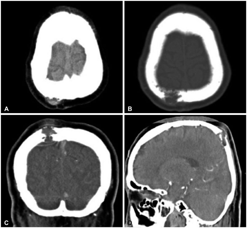
Fig. 1 Initial brain CT. CT scan (A) shows a right-sided superior parietal mass expanding the calvaria. Bone window CT (B) demonstrates a bony destruction. C and D reveal a well-defined, enhancing soft tissue mass in right parietal bone.
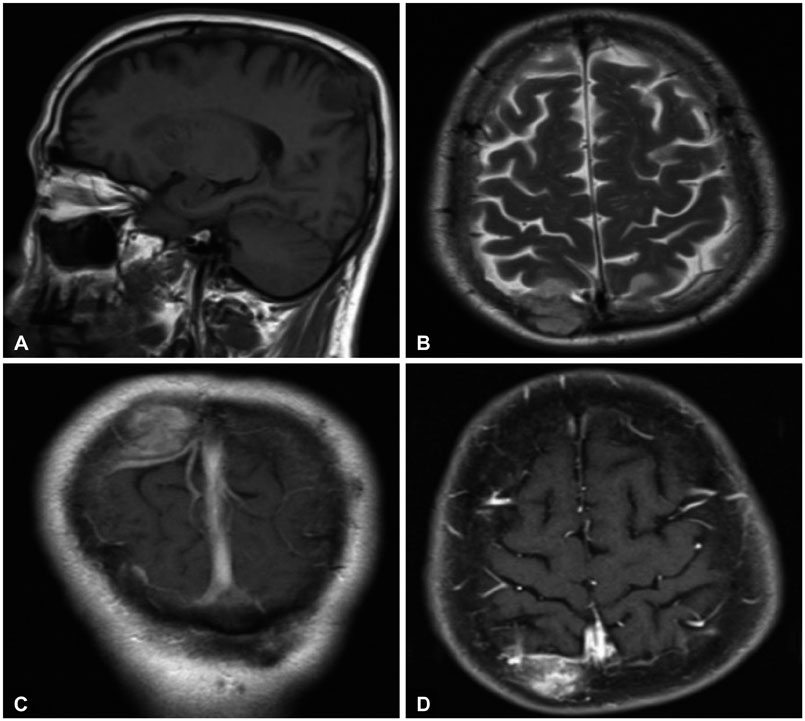
Fig. 2 Preoperative magnetic resonance images. T1 saggital weighted (A) MR shows a round shape, scalp bulging, iso-signal intensity. No parenchymal invasion on T2 axial weighted (B) image. Postcontrast T1-weighted MR (C and D) reveal a well-defined, lobulated, enhanced round mass in the right parietal bone (2.06×2.8 cm), with bone destruction and an intracranial extension compressing the brain.
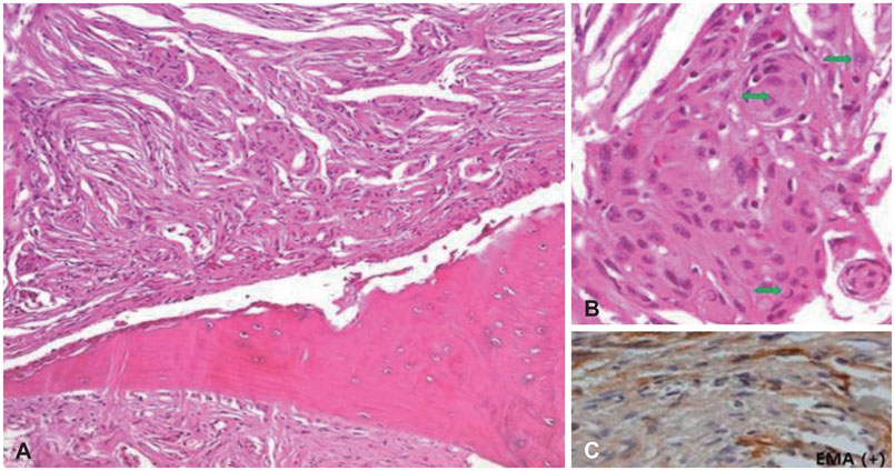
Fig. 3 Pathologic findings. A: The tumor cells are growing in a whorling pattern type (H&E, ×100). B: The tumor cells display typical subnuclear inclusions (H&E, ×200) (green arrows). C: The tumor cells express epithelial membrane antigen (EMA), which strongly suggests meningothelial differentiation. H&E, hematoxylin and eosin.
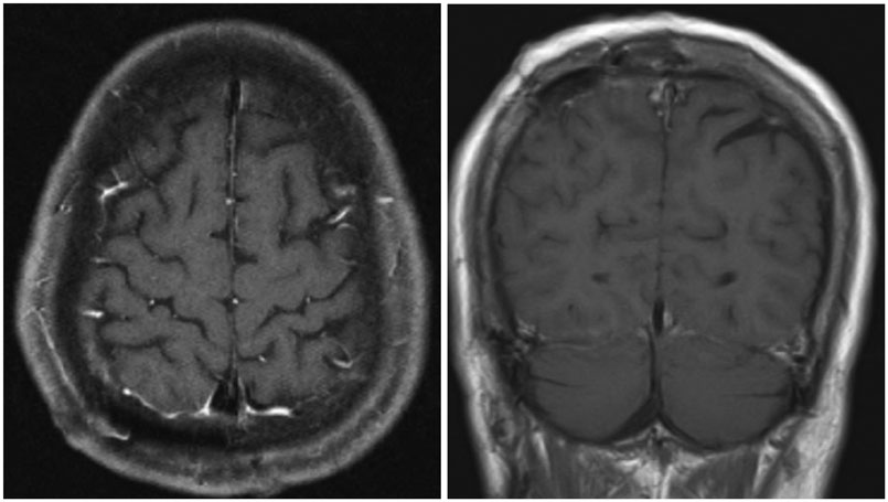
Fig. 4 Postoperative images show that was no evidence of residual mass at operative site.

Fig. 5 Comparative CT scans. Both A and B have osteolytic skull lesions at right parietal area. A: The tumor has a broad calvarial base, more expansion of the soft tissue. B: The tumor has a broad dural base, brain edema and more expansion of the brain parenchyme.
Reference
-
1. Craig WM, Gogela LJ. Intraorbital meningiomas; a clinicopathologic study. Am J Ophthalmol. 1949; 32:1663–1680. illust.
Article2. Agrawal V, Ludwig N, Agrawal A, Bulsara KR. Intraosseous intracranial meningioma. AJNR Am J Neuroradiol. 2007; 28:314–315.3. Matschke J, Addo J, Bernreuther C, Zustin J. Osseous changes in meningioma en plaque. Anticancer Res. 2011; 31:591–596.4. Elder JB, Atkinson R, Zee CS, Chen TC. Primary intraosseous meningioma. Neurosurg Focus. 2007; 23:E13.
Article5. Lang FF, Macdonald OK, Fuller GN, DeMonte F. Primary extradural meningiomas: a report on nine cases and review of the literature from the era of computerized tomography scanning. J Neurosurg. 2000; 93:940–950.
Article6. Arana E, Menor F, Lloret RM. Intraosseous meningioma. J Neurosurg. 1996; 85:362–363.7. Halpin SF, Britton J, Wilkins P, Uttley D. Intradiploic meningiomas. A radiological study of two cases confirmed histologically. Neuroradiology. 1991; 33:247–250.8. Azar-Kia B, Sarwar M, Marc JA, Schechter MM. Intraosseous meningioma. Neuroradiology. 1974; 6:246–253.
Article9. Yun JH, Lee SK. Primary osteolytic intraosseous atypical meningioma with soft tissue and dural invasion: report of a case and review of literatures. J Korean Neurosurg Soc. 2014; 56:509–512.
Article10. Turner OA, Laird AT. Meningioma with traumatic etiology. Report of a case. J Neurosurg. 1966; 24:96–98.11. Crawford TS, Kleinschmidt-DeMasters BK, Lillehei KO. Primary intraosseous meningioma. Case report. J Neurosurg. 1995; 83:912–915.12. Daffner RH, Yakulis R, Maroon JC. Intraosseous meningioma. Skeletal Radiol. 1998; 27:108–111.
Article13. Gibson SE, Prayson RA. Primary skull lesions in the pediatric population: a 25-year experience. Arch Pathol Lab Med. 2007; 131:761–766.
Article14. Stark AM, Eichmann T, Mehdorn HM. Skull metastases: clinical features, differential diagnosis, and review of the literature. Surg Neurol. 2003; 60:219–225. discussion 225-6.
Article15. Ghobashy A, Tobler W. Intraosseous calvarial meningioma of the skull presenting as a solitary osteolytic skull lesion: case report and review of the literature. Acta Neurochir (Wien). 1994; 129:105–108.
Article16. Hećimović I, Dmitrović B, Rubin O, Rukovanjski M, Vranković D. Skull osteolysis after mild head trauma: case report. Surg Neurol. 1999; 51:43–46.
Article
- Full Text Links
-
- Actions
-
Cited
- CITED
-
- Close
- Share
-
- Similar articles
-
- Primary Osteolytic Intraosseous Atypical Meningioma with Soft Tissue and Dural Invasion: Report of a Case and Review of Literatures
- Primary Intraosseous Calvarial Meningioma: A Case Report
- Two Cases of Primary Osteolytic Intraosseous Meningioma of the Skull Metastasizing to Whole Skull and the Spine
- Ewing's Sarcoma Mimicking a Meningioma in Radiological Findings: A Case Report
- Intradiploic Meningioma Presenting as on Osteolytic Skull Lesion Case Report
