Brainstem Congestion due to Dural Ateriovenous Fistula at the Craniocervical Junction
- Affiliations
-
- 1Department of Neurosurgery, Jinling Hospital, School of Medicine, Nanjing University, Nanjing, China. zhangxsp@163.com
- 2Department of Neurosurgery, Seoul St. Mary's Hospital, The Catholic University of Korea College of Medicine, Seoul, Korea.
- KMID: 2191081
- DOI: http://doi.org/10.3340/jkns.2014.55.3.152
Abstract
- Dural ateriovenous fistula (DAVF) at the craniocervical junction is rare. We report a patient presenting with brainstem dysfunction as an uncommon onset. Brainstem lesion was suggested by magnetic resonance image study. Angiogram revealed a DAVF at a high cervical segment supplied by the meningeal branch of the right vertebral artery, with ascending and descending venous drainage. Complete obliteration of the fistula was achieved via transarterial Onyx embolization. Clinical cure was achieved in the follow-up period; meanwhile, imaging abnormalities of this case disappeared. Accordingly, we hypothesize that a brainstem lesion of this case was caused by craniocervical DAVF, which induced venous hypertension. Thus, venous drainage patterns should be paid attention to because they are important for diagnosis and theraputic strategy.
MeSH Terms
Figure
-
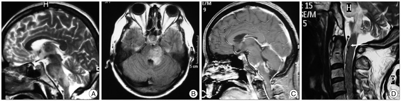
Fig. 1 Sagittal T2-weight MRI (A) and FLAIR image (B) demonstrating abnormalities from pons to medulla oblongata. C : Sagittal contrast enhanced imaging demonstrating abnormal enhancing lesions located at pontobulbar junction, dorsal oblongata and craniocervical junction. D : Sagittal cervical T2-weight MRI demonstrating abnormal flow void (arrow) at ventral surface from craniocervical junction to cervical cord.
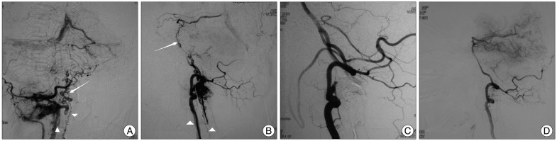
Fig. 2 A and B : Right VA angiogram, posterior-anterior and lateral view, demonstrating DAVF with a meningeal branch originated from radicular artery of the right C2 segment of VA as a feeding vessel, draining via abnormally hypertrophic pontomesencephalic veins (arrows) ascending from basal vein retrogradely into straight sinus and descending into the anterior spinal vein, anterior internal vertebral venous plexus and vertebral artery venous plexus (arrowheads). C : Right VA angiogram immediately after embolization, lateral view, demonstrating complete obliteration of the fistula. D : Right VA angiogram six months after embolization, lateral view, demonstrating complete obliteration of the fistula and disappearance of abnormal venous drainage. VA : vertebral artery, DAVF : dural ateriovenous fistula.
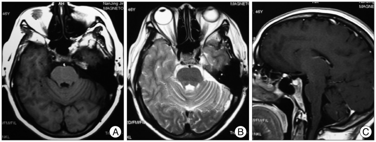
Fig. 3 Follow-up six-month brain MRI. Axial T1-weight imaging (A), axial T2-weight imaging (B), and contrast enhanced T1-weight imaging (C) demonstrating disappeared brainstem abnormal signals.
Reference
-
1. Bussière M, Lownie SP, Pelz DM, Nicolle D. Direct carotid-cavernous fistula causing brainstem venous congestion. J Neuroophthalmol. 2009; 29:21–25. PMID: 19458571.
Article2. Chen G, Wang Q, Tian Y, Gu Y, Xu B, Leng B, et al. Dural arteriovenous fistulae at the craniocervical junction : the relation between clinical symptom and pattern of venous drainage. Acta Neurochir Suppl. 2011; 110(Pt 2):99–104. PMID: 21125453.3. Fassett DR, Rammos SK, Patel P, Parikh H, Couldwell WT. Intracranial subarachnoid hemorrhage resulting from cervical spine dural arteriovenous fistulas : literature review and case presentation. Neurosurg Focus. 2009; 26:E4. PMID: 19119890.4. Guo LM, Zhou HY, Xu JW, Wang GS, Tian X, Wang Y, et al. Dural arteriovenous fistula at the foramen magnum presenting with subarachnoid hemorrhage : case reports and literature review. Eur J Neurol. 2010; 17:684–691. PMID: 20050886.
Article5. Hamada J, Yano S, Kai Y, Koga K, Morioka M, Ishimaru Y, et al. Histopathological study of venous aneurysms in patients with dural arteriovenous fistulas. J Neurosurg. 2000; 92:1023–1027. PMID: 10839265.
Article6. Iwasaki M, Murakami K, Tomita T, Numagami Y, Nishijima M. Cavernous sinus dural arteriovenous fistula complicated by pontine venous congestion. A case report. Surg Neurol. 2006; 65:516–518. discussion 519. PMID: 16630921.
Article7. Jellema K, Tijssen CC, van Gijn J. Spinal dural arteriovenous fistulas : a congestive myelopathy that initially mimics a peripheral nerve disorder. Brain. 2006; 129(Pt 12):3150–3164. PMID: 16921175.
Article8. Kai Y, Hamada J, Morioka M, Yano S, Mizuno T, Kuratsu J. Arteriovenous fistulas at the cervicomedullary junction presenting with subarachnoid hemorrhage : six case reports with special reference to the angiographic pattern of venous drainage. AJNR Am J Neuroradiol. 2005; 26:1949–1954. PMID: 16155140.9. Kai Y, Hamada JI, Morioka M, Yano S, Ushio Y. Brain stem venous congestion due to dural arteriovenous fistulas of the cavernous sinus. Acta Neurochir (Wien). 2004; 146:1107–1111. discussion 1111-1112. PMID: 15744846.
Article10. Kulwin C, Bohnstedt BN, Scott JA, Cohen-Gadol A. Dural arteriovenous fistulas presenting with brainstem dysfunction : diagnosis and surgical treatment. Neurosurg Focus. 2012; 32:E10. PMID: 22537119.11. Lin CH, Lo YK, Lin YT, Li JY, Lai PH, Gau YY. Central vasomotor failure in a patient with medulla arteriovenous fistula. Acta Neurol Taiwan. 2006; 15:192–196. PMID: 16995599.12. Murata H, Kubota T, Murai M, Kanno H, Fujii S, Yamamoto I. Brainstem congestion caused by direct carotid-cavernous fistula--case report. Neurol Med Chir (Tokyo). 2003; 43:255–258. PMID: 12790286.
Article13. Park KW, Park SI, Im SB, Kim BT. Spinal dural arteriovenous fistula with supply from the lateral sacral artery-case report and review of literature-. J Korean Neurosurg Soc. 2009; 45:115–117. PMID: 19274124.
Article14. Rhoton AL Jr. The posterior fossa veins. Neurosurgery. 2000; 47(3 Suppl):S69–S92. PMID: 10983305.
Article15. Shintani S, Tsuruoka S, Shiigai T. Carotid-cavernous fistula with brainstem congestion mimicking tumor on MRI. Neurology. 2000; 55:1929–1931. PMID: 11134402.
Article16. Stiefel MF, Albuquerque FC, Park MS, Dashti SR, McDougall CG. Endovascular treatment of intracranial dural arteriovenous fistulae using Onyx : a case series. Neurosurgery. 2009; 65(6 Suppl):132–139. discussion 139-140. PMID: 19934987.17. Takahashi S, Tomura N, Watarai J, Mizoi K, Manabe H. Dural arteriovenous fistula of the cavernous sinus with venous congestion of the brain stem : report of two cases. AJNR Am J Neuroradiol. 1999; 20:886–888. PMID: 10369361.18. Teng MM, Chang T, Pan DH, Chang CN, Huang CI, Guo WY, et al. Brainstem edema : an unusual complication of carotid cavernous fistula. AJNR Am J Neuroradiol. 1991; 12:139–142. PMID: 1899502.19. Terao T, Taniguchi M, Ide K, Shinozaki M, Takahashi H. Cervical dural arteriovenous fistula presenting with brainstem dysfunction : case report and review. Spine (Phila Pa 1976). 2006; 31:E722–E727. PMID: 16946647.
- Full Text Links
-
- Actions
-
Cited
- CITED
-
- Close
- Share
-
- Similar articles
-
- Dural Arteriovenous Fistula Presenting as Acute Unilateral Vestibulopathy
- Ruptured Medullary Hemangioblastoma Mimicking a Craniocervical Junction Dural Arteriovenous Fistula with a Pseudoaneurysm
- Arteriovenous Fistula at the Craniocervical Junction Found After Cervical Laminoplasty for Ossification of the Posterior Longitudinal Ligament
- Treatment of a Traumatic Carotid Cavernous Fistula and Dural Shunt Using Microcoils
- Anterior Craniocervical Junctional Neurenteric Cyst
