Malignant Transformation of an Epidermoid Cyst in the Cerebellopontine Angle
- Affiliations
-
- 1Department of Neurosurgery, Chonbuk National University Hospital and Medical School, Jeonju, Korea. nsjmlee@gmail.com
- KMID: 2190555
- DOI: http://doi.org/10.3340/jkns.2012.52.2.148
Abstract
- Intracranial squamous cell carcinoma is extremely rare, with most of the cases arising from malignant transformation of an epidermoid or a dermoid cyst. The patient presented with facial weakness. Initial magnetic resonance imaging revealed a mass in the right cerebellopontine angle. A subtotal resection was performed via right retrosigmoid suboccipital approach. Histopathological findings were consistent with an epidermoid tumor. Five months later, the patient underwent gamma knife radiosurgery due to highly probable recurrent epidermoid tumor. Two years after, the patient's neurological deficit had been newly developed, and follow-up magnetic resonance imaging demonstrated a large contrast-enhancing tumor in the left cerebellopontine angle, which compressed the brainstem. After resection of the tumor, histopathological examinations revealed a squamous cell carcinoma probably arising from an underlying epidermoid cyst. We report a case of an epidermoid tumor in the cerebellopontine angle that transformed into a squamous cell carcinoma.
Keyword
MeSH Terms
Figure
-

Fig. 1 A : T2-weighted image shows a high signal mass in the right CPA. B : Contrast-enhanced T1-weighted image shows nonenhancing huge mass. C : Diffusion weighted image shows very high signal intensity mass in the right CPA suggestive of an epidermoid cyst. D : The cyst is lined by keratinizing squamous epithelium and filled with lamellated keratinous debris (Hematoxylin-Eosin stain, ×100). CPA : cerebellopontine angle.
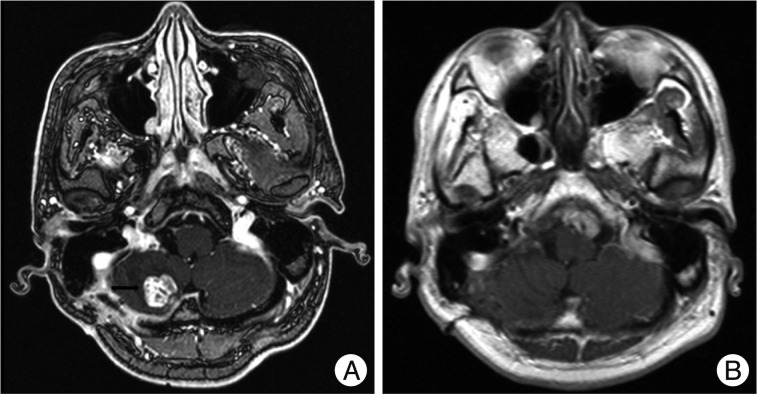
Fig. 2 A : In July 2009, contrast-enhanced axial T1-weighted image shows strong enhancement of the small nodular lesion (black arrow) in the right cerebellar hemisphere. The patient received GKRS in this lesion. B : In January 2011, follow-up contrast-enhanced axial T1-weighted image shows that there is no enhanced tumor. GKRS : gamma knife radiosurgery.

Fig. 3 A : T2-weighted image shows isodense signal intensity mass in the left CPA. B : Contrast-enhanced axial T1-weighted image shows strong enhancement of the nodular lesion in the left CPA. C : Contrast-enhanced sagittal T1-weighted image shows strong enhancement. D : The specimen contains high cellular area with enlarged pleomorphic and hyperchromatic nuclei, which is a component of poorly differentiated squamous cell carcinoma (Hematoxylin-Eosin stain, ×200). CPA : cerebellopontine angle.
Reference
-
1. Abramson RC, Morawetz RB, Schlitt M. Multiple complications from an intracranial epidermoid cyst : case report and literature review. Neurosurgery. 1989; 24:574–578. PMID: 2651960.
Article2. Chen S, Ikawa F, Kurisu K, Arita K, Takaba J, Kanou Y. Quantitative MR evaluation of intracranial epidermoid tumors by fast fluid-attenuated inversion recovery imaging and echo-planar diffusion-weighted imaging. AJNR Am J Neuroradiol. 2001; 22:1089–1096. PMID: 11415903.3. Cheng MH, Lin JW. Intracranial meningioma with intratumoral hemorrhage. J Formos Med Assoc. 1997; 96:116–120. PMID: 9071837.4. deSouza CE, deSouza R, da Costa S, Sperling N, Yoon TH, Abdelhamid MM, et al. Cerebellopontine angle epidermoid cysts : a report on 30 cases. J Neurol Neurosurg Psychiatry. 1989; 52:986–990. PMID: 2795068.5. Dunn RC Jr, Archer CA, Rapport RL 2nd, Looi LM. Unusual CT-dense posterior fossa epidermoid cyst : case report. J Neurosurg. 1981; 55:654–656. PMID: 7277016.6. Gao PY, Osborn AG, Smirniotopoulos JG, Harris CP. Radiologic-pathologic correlation. Epidermoid tumor of the cerebellopontine angle. AJNR Am J Neuroradiol. 1992; 13:863–872. PMID: 1590184.7. Garcia CA, McGarry PA, Rodriguez F. Primary intracranial squamous cell carcinoma of the right cerebellopontine angle. J Neurosurg. 1981; 54:824–828. PMID: 7017078.
Article8. Guidetti B, Gagliardi FM. Epidermoid and dermoid cysts. Clinical evaluation and late surgical results. J Neurosurg. 1977; 47:12–18. PMID: 864501.9. Hamlat A, Hua ZF, Saikali S, Laurent JF, Gedouin D, Ben-Hassel M, et al. Malignant transformation of intra-cranial epithelial cysts : systematic article review. J Neurooncol. 2005; 74:187–194. PMID: 16193391.
Article10. Kida Y, Yoshimoto M, Hasegawa T, Fujitani S. [Radiosurgery of epidermoid tumors with gamma knife : possiblility of radiosurgical nerve decompression]. No Shinkei Geka. 2006; 34:375–381. PMID: 16613218.11. Link MJ, Cohen PL, Breneman JC, Tew JM Jr. Malignant squamous degeneration of a cerebellopontine angle epidermoid tumor. Case report. J Neurosurg. 2002; 97:1237–1243. PMID: 12450053.
Article12. Mayayo Vicente MS, Fernández Arjona M, Gascón Veguín JP, Jiménez Sánchez F, Puig Rullan AM, Cortés Arónguéz I. [Prostatic epidermoid carcinoma : report of a new case an review of the literature]. Arch Esp Urol. 2003; 56:939–943. PMID: 14639850.13. Mori Y, Suzuki Y, Tanasawa T, Yoshida J, Wakabayashi T, Kobayashi T. [A case report of epidermoid carcinoma in the cerebello-pontine angle]. No Shinkei Geka. 1995; 23:905–909. PMID: 7477700.14. Nagasawa D, Yew A, Spasic M, Choy W, Gopen Q, Yang I. Survival outcomes for radiotherapy treatment of epidermoid tumors with malignant transformation. J Clin Neurosci. 2012; 19:21–26. PMID: 22024232.
Article15. Sawan B, Vital A, Loiseau H, Dousset V, Strub D, Vital C. Squamous cell carcinoma developing in an intracranial prepontine epidermoid cyst. Ann Pathol. 2000; 20:258–260. PMID: 10891726.
- Full Text Links
-
- Actions
-
Cited
- CITED
-
- Close
- Share
-
- Similar articles
-
- Primary Intracranial Epidermoid Carcinoma
- MR Imaging of a Cerebello-Pontine Angle Epidermoid Cyst with a Malignant Transformation: Case Report
- Isolated Trigeminal Neuralgia Presented Due to Cerebellopontine Angle Epidermoid Cyst
- A Case of Epidermoid Cyst in the Fourth Ventricle
- Primary Intracranial Squamous Cell Carcinoma in the Brain Stem with a Cerebellopontine Angle Epidermoid Cyst
