Spinal Intramedullary Ependymal Cysts : A Case Report and Review of the Literature
- Affiliations
-
- 1Department of Neurosurgery, Seoul National University Hospital, Seoul National University College of Medicine, Seoul, Korea.
- 2Department of Neurosurgery, Seoul National University Bundang Hospital, Seoul National University College of Medicine, Seongnam, Korea. jibkim@snu.ac.kr
- KMID: 2190528
- DOI: http://doi.org/10.3340/jkns.2012.52.1.67
Abstract
- We report a rare case of a spinal intramedullary ependymal cyst in a 46-year-old female and review the 17 pathologically proven cases in the literature. The patient presented with a two-week history of gradually increasing tingling in her left posterior thigh and calf. A preoperative magnetic resonance image revealed a well-defined intramedullary cystic lesion on the ventral side of the spinal cord at the T11 to T12 levels. The lesion was hyper intense in T2-weighted images and hypointense in T1-weighted. The patient underwent a right-side hemilaminectomy at the T11 to T12 levels and fenestration of the cyst wall. After having the cyst wall partially removed and communication established between the cyst and the subarachnoid space, the patient improved neurologically. A histological study of the surgical specimens revealed that the cyst wall consisted of glial cells lined by a simple cuboidal to columnar epithelium. An immunohistochemical examination of the cells lining the cyst wall was positive for S-100 protein, glial fibrillary acidic protein, epithelial membrane antigen, and cytokeratin. We suggest that the optimal treatment of intramedullary ependymal cysts creates adequate communication between the cyst and the subarachnoid space.
Keyword
MeSH Terms
Figure
-

Fig. 1 The magnetic resonance image revealing a well-defined cystic lesion on the ventral side of the spinal cord at the T11 to T12 levels. The lesion is a 36×15 mm oval intramedullary cystic mass. The lesion is hyperintense in T2-weighted images (A and C) and hypointense in T1-weighted images (B and D). In the axial imaging, the cyst is observed to compress the spinal cord on the left dorsal side (C and D).

Fig. 2 A glistening white cyst without hemorrhage or parenchyma (A). The cyst wall is fenestrated to obtain adequate communication between the cyst and the subarachnoid space (B).

Fig. 3 In the H&E staining (A), the cyst wall consists of glial cells lined by a simple cuboidal to columnar epithelium. An immunohistochemical examination of the cells lining the cyst wall is positive for glial fibrillary acidic protein (B), S-100 protein (C), and cytokeratin (D). These findings are consistent with an ependymal cyst diagnosis.
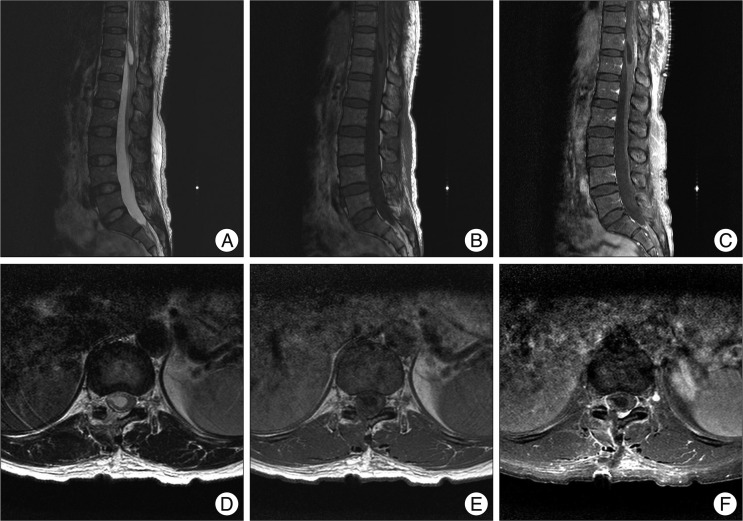
Fig. 4 A postoperative magnetic resonance image reveals that the cystic lesion's size had decreased to 29×9.6 mm and that the spinal cord had decompressed.
Reference
-
1. Chhabra R, Bansal S, Radotra BD, Mathuriya SN. Recurrent intramedullary cervical ependymal cyst. Neurol India. 2003; 51:111–113. PMID: 12865539.2. Dharker SR, Kanhere S, Dharker RS. Intramedullary epithelial cyst of the spinal cord. Surg Neurol. 1979; 12:443–444. PMID: 524259.3. Findler G, Hadani M, Tadmor R, Bubis JJ, Shaked I, Sahar A. Spinal intradural ependymal cyst: a case report and review of the literature. Neurosurgery. 1985; 17:484–486. PMID: 4047361.
Article4. Fortuna A, Mercuri S. Intradural spinal cysts. Acta Neurochir (Wien). 1983; 68:289–314. PMID: 6880882.
Article5. Iwahashi H, Kawai S, Watabe Y, Chitoku S, Akita N, Fuji T, et al. Spinal intramedullary ependymal cyst: a case report. Surg Neurol. 1999; 52:357–361. PMID: 10555841.
Article6. Kato M, Nakamura H, Suzuki E, Terai H, Wakasa K, Wakasa T, et al. Ependymal cyst in the lumbar spine associated with cauda equina compression. J Clin Neurosci. 2008; 15:827–830. PMID: 18407500.
Article7. Kumar R, Nayak SR, Krishnani N, Chhabra DK. Spinal intramedullary ependymal cyst. Report of two cases and review of the literature. Pediatr Neurosurg. 2001; 35:29–34. PMID: 11490188.8. Lalitha AV, Rout P, D Souza F, Shailesh , Rao S. Spinal intramedullary neuroepithelial (ependymal) cyst. A rare cause of treatable acute para paresis. Indian J Pediatr. 2006; 73:945–946. PMID: 17090911.
Article9. Morest DK, Silver J. Precursors of neurons, neuroglia, and ependymal cells in the CNS : what are they? Where are they from? How do they get where they are going? Glia. 2003; 43:6–18. PMID: 12761861.
Article10. Nagano S, Ijiri K, Kawabata R, Zenmyo M, Yone K, Kitajima S, et al. Ependymal cyst in the conus medullaris. J Clin Neurosci. 2010; 17:272–273. PMID: 20036544.
Article11. Pagni CA, Canavero S, Vinattieri A, Forni M. Intramedullary spinal ependymal cyst: case report. Surg Neurol. 1991; 35:325–328. PMID: 2008649.
Article12. Robertson DP, Kirkpatrick JB, Harper RL, Mawad ME. Spinal intramedullary ependymal cyst. Report of three cases. J Neurosurg. 1991; 75:312–316. PMID: 2072172.13. Rousseau M, Lesoin F, Combelles G, Krivosic Y, Warot P. An intramedullary ependymal cyst in a 71-year-old woman. Neurosurgery. 1983; 13:52–54. PMID: 6877566.
Article14. Saito K, Morita A, Shibahara J, Kirino T. Spinal intramedullary ependymal cyst: a case report and review of the literature. Acta Neurochir (Wien). 2005; 147:443–446. discussion 446. PMID: 15666187.
Article15. Sharma BS, Banerjee AK, Khosla VK, Kak VK. Congenital intramedullary spinal ependymal cyst. Surg Neurol. 1987; 27:476–480. PMID: 3563862.
Article16. Takci E, Sengul G, Keles M. Spinal intramedullary ependymal cyst and tethered cord in an adult. Case report. J Neurosurg Spine. 2006; 4:506–508. PMID: 16776364.
