Solitary Xanthogranuloma of the Upper Cervical Spine in a Male Adult
- Affiliations
-
- 1Department Neurosurgery, Kyung Hee University Hospital at Gangdong, Seoul, Korea. apuzzo@hanmail.net
- KMID: 2190475
- DOI: http://doi.org/10.3340/jkns.2012.51.1.54
Abstract
- We present the rare case of solitary xanthogranuloma in the upper cervical column mimicking a Brown-Sequard syndrome. A 29-year-old man complained with right hemiparesis and left hypoesthesia after a car accident. Computed tomography and magnetic resonance images revealed a lobulated homogenously well-enhancing mass in between posterior arch of the atlas (C1) and spinous process of the axis (C2) resulting in a marked spinal canal narrowing with cortical erosions. The patient was managed by complete resection of the tumor with partial laminectomy with lower half of C1 posterior arch and upper half of C2 spinous process. The authors advise complete removal of the xanthogranuloma and consideration as a differential diagnosis of lesions among upper cervical lesions.
MeSH Terms
Figure
-
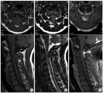
Fig. 1 Preoperative MRI showing abnormal signal intensities on epidural dumbbell-shaped mass (measuring 25×18×24 mm) traversing the C1 to C2 interspinous space, compressing surrounding structures. The dumbbell-shaped lesion reveals isointense on sagittal T1-WI (A), mixed hypointense on T2-WI (B), and well-enhanced after gadolinium administration (C). The spinal cord was compressed at the C2 level, and the signal change appeared on T2-WI. MRI : magnetic resonance images, WI : weighted image.
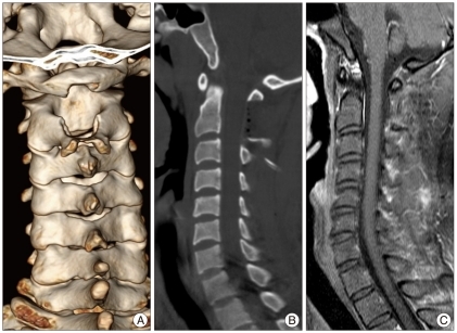
Fig. 2 Post-operative CT scan. Partial hemilaminectomy, from the lower half of the C1 posterior arch to upper half of C2 spinous process, was performed (A : mid-sagittal, B : 3-dimensional reconstruction). C : Follow-up MRI with enhancement after postoperative 2 years reveals no residual and no recurrence. CT : computed tomography, MRI : magnetic resonance images.

Fig. 3 Photomicrographs of the tumor reveals focal aggregation of cells with ample, clear, and foamy cytoplasm. There are large round cells with irregular vesicular nuclei. H&E, original magnification ×200.

Fig. 4 Gross finding of the tumor. A : External surface of the tumor; ovoid, yellowish, encapsulated with a white-colored adhesion scar at the mid-portion of the mass shown in the picture. B : Mid-section of the tumor, particularly yellow and white with cystic components.
Reference
-
1. Adamson NF. Congenital xanthoma multiplex in a child. Br J Dermatol. 1905; 17:222–223.2. Agabegi SS, Iorio TE, Wilson JD, Fischgrund JS. Juvenile xanthogranuloma in an adult lumbar spine : a case report. Spine (Phila Pa 1976). 2011; 36:E69–E73. PMID: 21192217.3. Ayres WW, Haymaker W. Xanthoma and cholesterol granuloma of the choroid plexus. Report of the pathological aspects in 29 cases. J Neuropathol Exp Neurol. 1960; 19:280–295. PMID: 13795372.
Article4. Boström J, Janssen G, Messing-Jünger M, Felsberg JU, Neuen-Jacob E, Engelbrecht V, et al. Multiple intracranial juvenile xanthogranulomas. Case report. J Neurosurg. 2000; 93:335–341.5. Cao D, Ma J, Yang X, Xiao J. Solitary juvenile xanthogranuloma in the upper cervical spine : case report and review of the literatures. Eur Spine J. 2008; 17(Suppl 2):S318–S323. PMID: 18228052.6. Castro-Gago M, Gómez-Lado C, Alvez F, Alonso A, Vieites B. Juvenile xanthogranuloma of the cauda equina. Pediatr Neurol. 2009; 40:123–125. PMID: 19135628.
Article7. Dehner LP. Juvenile xanthogranulomas in the first two decades of life : a clinicopathologic study of 174 cases with cutaneous and extracutaneous manifestations. Am J Surg Pathol. 2003; 27:579–593. PMID: 12717244.
Article8. Freyer DR, Kennedy R, Bostrom BC, Kohut G, Dehner LP. Juvenile xanthogranuloma : forms of systemic disease and their clinical implications. J Pediatr. 1996; 129:227–237. PMID: 8765620.
Article9. George DH, Scheithauer BW, Hilton DL, Fakhouri AJ, Kraus EW. Juvenile xanthogranuloma of peripheral nerve : a report of two cases. Am J Surg Pathol. 2001; 25:521–526. PMID: 11257628.10. Hernandez-Martin A, Baselga E, Drolet BA, Esterly NB. Juvenile xanthogranuloma. J Am Acad Dermatol. 1997; 36:355–367. quiz 368-369. PMID: 9091465.
Article11. Inoue H, Seichi A, Yamamuro K, Kojima M, Kimura A, Hoshino Y. Dumbbell-type juvenile xanthogranuloma in the cervical spine of an adult. Eur Spine J. 2011; 20(Suppl 2):S343–S347. PMID: 21468645.
Article12. Iwasaki Y, Hida K, Nagashima K. Cauda equina xanthogranulomatosis. Br J Neurosurg. 2001; 15:72–73. PMID: 11303669.
Article13. Jain A, Mathur K, Khatri S, Kasana S, Jain SK. Rare presentation of juvenile xanthogranuloma in the thoracic spine of an adult patient : case report and literature review. Acta Neurochir (Wien). 2011; 153:1813–1818. PMID: 21626171.
Article14. Kim DS, Kim TS, Choi JU. Intradural extramedullary xanthoma of the spine : a rare lesion arising from the dura mater of the spine : case report. Neurosurgery. 1996; 39:182–185. PMID: 8805158.
Article15. Kitchen ND, Davies MS, Taylor W. Juvenile xanthogranuloma of nerve root origin. Br J Neurosurg. 1995; 9:233–237. PMID: 7632374.
Article16. Kraus MD, Haley JC, Ruiz R, Essary L, Moran CA, Fletcher CD. "Juvenile" xanthogranuloma : an immunophenotypic study with a reappraisal of histogenesis. Am J Dermatopathol. 2001; 23:104–111. PMID: 11285404.17. Lesniak MS, Viglione MP, Weingart J. Multicentric parenchymal xanthogranuloma in a child : case report and review of the literature. Neurosurgery. 2002; 51:1493–1498. discussion 1498. PMID: 12445357.
Article18. Paulus W, Kirchner T, Michaela M, Kühl J, Warmuth-Metz M, Sörensen N, et al. Histiocytic tumor of Meckel's cave. An intracranial equivalent of juvenile xanthogranuloma of the skin. Am J Surg Pathol. 1992; 16:76–83. PMID: 1728198.19. Rampini PM, Alimehmeti RH, Egidi MG, Zavanone ML, Bauer D, Fossali E, et al. Isolated cervical juvenile xanthogranuloma in childhood. Spine (Phila Pa 1976). 2001; 26:1392–1395. PMID: 11426158.
Article20. Schultz KD Jr, Petronio J, Narad C, Hunter SB. Solitary intracerebral juvenile xanthogranuloma. Case report and review of the literature. Pediatr Neurosurg. 1997; 26:315–321. PMID: 9485160.21. Shimosawa S, Tohyama K, Shibayama M, Takeuchi H, Hirota T. Spinal xanthogranuloma in a child : case report. Surg Neurol. 1993; 39:138–142. PMID: 8351627.
