Expression of cytokeratin 10, 16 and 17 as biomarkers differentiating odontogenic keratocysts from dentigerous cysts
- Affiliations
-
- 1Department of Oral and Maxillofacial Surgery, School of Dentistry, Kyungpook National University, Daegu, Korea. kimcs@knu.ac.kr
- KMID: 2189694
- DOI: http://doi.org/10.5125/jkaoms.2012.38.2.78
Abstract
OBJECTIVES
Odontogenic keratocysts (OKCs) have a tendency to recur and possess an aggressive nature. the aim of the present study was to evaluate cytokeratin (CK) expression patterns as a method for the differentiation between dentigerous cysts (DCs) and OKCs, as their histomorphologic appearance are often indistinguishable.
MATERIALS AND METHODS
Formalin-fixed, paraffin-embedded tissue sections of 43 OKCs and 38 DCs were immunohistochemically analyzed with i-solution in a quantitative manner in order to evaluate the immunoreactivity of CK 10, 16 and 17.
RESULTS
CK 10 expression was evident in 79.1% of OKCs but found in only 18.4% of DCs (P<0.05), and CK 10 expression was observed to occur more frequently in OKCs (mean 25.45%) than in DCs (2.19%) (P<0.05). The expression of CK 16 was evident in 79.1% of OKCs but found in only 7.9% of the DCs (P<0.05) and CK 16 expression was observed to occur more frequently in OKCs (mean 4.33%) than in the DCs (0.61%) (P<0.05). The expression of CK 17 was evident in 88.4% of OKCs but seen in only 15.7% of the DCs (P<0.05) and CK 17 expression was observed to occur more frequently in OKCs (mean 31.11%) than in the DCs (2.37%) (P<0.05).
CONCLUSION
The immunohistochemical detection of CK 10, 16 and 17 can be utilized as a valuable biomarker for use in distinguishing between OKCs and DCs, which have clinically significant differential diagnoses.
MeSH Terms
Figure
-

Fig. 1 Photomicrograph of the epithelium of odontogenic keratocyst. The maximum intensity of the immunohistochemical expression of cytokeratin (CK) 10 and basal and suprabasal cells were strongly positive for CK 10 (H&E staining, ×40).

Fig. 2 Photomicrograph of the epithelium of the dentigerous cyst. The mean intensity of the immunohistochemical expression of cytokeratin (CK) 10 and basal cells were weakly positive for CK 10 (H&E staining, ×40).

Fig. 3 Photomicrograph of the epithelium of odontogenic keratocyst. The maximum intensity of the immunohistochemical expression of cytokeratin (CK) 16 and basal and suprabasal cells were strongly positive for CK 16 (H&E staining, ×40).
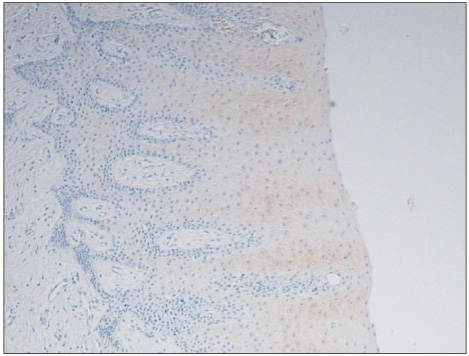
Fig. 4 Photomicrograph of the epithelium of the dentigerous cyst. The mean intensity of the immunohistochemical expression of cytokeratin (CK) 16 and basal cells were weakly positive for CK 16 (H&E staining, ×40).
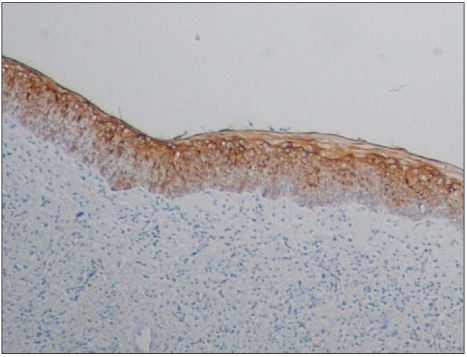
Fig. 5 Photomicrograph of the epithelium of odontogenic keratocyst. The maximum intensity of the immunohistochemical expression of cytokeratin (CK) 17 and basal and suprabasal cells were strongly positive for CK 17 (H&E staining, ×40).

Fig. 6 Photomicrograph of the epithelium of the dentigerous cyst. The mean intensity of the immunohistochemical expression of cytokeratin (CK) 17 and basal cells were almost negative for CK 17 (H&E staining, ×40).
Reference
-
1. Philipsen HP. Om keratocyster (kolesteatomer) i kaeberne. Tandlaegebladet. 1956. 60:963–980.2. Pindborg JJ, Hansen J. Studies on odontogenic cyst epithelium: 2. Clinical and roentgenologic aspects of odontogenic keratocysts. Acta Pathol Microbiol Scand. 1963. 58:283–294.3. Barnes L. Surgical pathology of the head and neck. 1986. 1st ed. New York: Marcel Dekker Inc..4. el-Hajj G, Anneroth G. Odontogenic keratocysts--a retrospective clinical and histologic study. Int J Oral Maxillofac Surg. 1996. 25:124–129.
Article5. Chow HT. Odontogenic keratocyst: a clinical experience in Singapore. Oral Surg Oral Med Oral Pathol Oral Radiol Endod. 1998. 86:573–577.6. Shear M. The aggressive nature of the odontogenic keratocyst: is it a benign cystic neoplasm? Part 2. Proliferation and genetic studies. Oral Oncol. 2002. 38:323–331.7. Toller P. Origin and growth of cysts of the jaws. Ann R Coll Surg Engl. 1967. 40:306–336.8. Shear M. The aggressive nature of the odontogenic keratocyst: is it a benign cystic neoplasm? Part 1. Clinical and early experimental evidence of aggressive behaviour. Oral Oncol. 2002. 38:219–226.
Article9. Shear M. The aggressive nature of the odontogenic keratocyst: is it a benign cystic neoplasm? Part 3. Immunocytochemistry of cytokeratin and other epithelial cell markers. Oral Oncol. 2002. 38:407–415.
Article10. Li TJ, Kitano M, Chen XM, Itoh T, Kawashima K, Sugihara K, et al. Orthokeratinized odontogenic cyst: a clinicopathological and immunocytochemical study of 15 cases. Histopathology. 1998. 32:242–251.
Article11. Stoll C, Stollenwerk C, Riediger D, Mittermayer C, Alfer J. Cytokeratin expression patterns for distinction of odontogenic keratocysts from dentigerous and radicular cysts. J Oral Pathol Med. 2005. 34:558–564.
Article12. Philipsen HP. Barnes L, Eveson JW, Reichart P, Sidransky D, editors. Keratocystic odontogenic tumor. World Health Organization classification of tumors: pathology and genetics of head and neck tumors. 2005. Lyon: IARC Press;306–307.13. Taylor CR, Shi S-R, Barr NJ, Wu N. Dabbs DJ, editor. Techniques of immunohistochemistry. Diagnostic immunohistochemistry. 2002. Philadelphia, Pennsylvania: Churchill Livingstone;1–44.
Article14. Cerilli LA, Wick MR. Dabbs DJ, editor. Immunohistology of soft tissue and osseous neoplasm. Diagnostic immunohistochemistry. 2002. Philadelphia, Pennsylvania: Churchill Livingstone;59–112.15. Meara JG, Pilch BZ, Shah SS, Cunningham MJ. Cytokeratin expression in the odontogenic keratocyst. J Oral Maxillofac Surg. 2000. 58:862–865.
Article16. MacDonald AW, Fletcher A. Expression of cytokeratin in the epithelium of dentigerous cysts and odontogenic keratocysts: an aid to diagnosis. J Clin Pathol. 1989. 42:736–739.
Article17. August M, Faquin WC, Troulis M, Kaban LB. Differentiation of odontogenic keratocysts from nonkeratinizing cysts by use of fine-needle aspiration biopsy and cytokeratin-10 staining. J Oral Maxillofac Surg. 2000. 58:935–940.
Article18. Gao Z, Mackenzie IC, Cruchley AT, Williams DM, Leigh I, Lane EB. Cytokeratin expression of the odontogenic epithelia in dental follicles and developmental cysts. J Oral Pathol Med. 1989. 18:63–67.
Article19. Hormia M, Ylipaavalniemi P, Nagle RB, Virtanen I. Expression of cytokeratins in odontogenic jaw cysts: monoclonal antibodies reveal distinct variation between different cyst types. J Oral Pathol. 1987. 16:338–346.
Article20. Morgan PR, Shirlaw PJ, Johnson NW, Leigh IM, Lane EB. Potential applications of anti-keratin antibodies in oral diagnosis. J Oral Pathol. 1987. 16:212–222.
Article21. Gao Z, Mackenzie IC, Williams DM, Cruchley AT, Leigh I, Lane EB. Patterns of keratin-expression in rests of Malassez and periapical lesions. J Oral Pathol. 1988. 17:178–185.
Article22. Ahlfors E, Larsson A, Sjögren S. The odontogenic keratocyst: a benign cystic tumor? J Oral Maxillofac Surg. 1984. 42:10–19.
Article23. Vedtofte P, Praetorius F. Recurrence of the odontogenic keratocyst in relation to clinical and histological features. A 20-year follow-up study of 72 patients. Int J Oral Surg. 1979. 8:412–420.
Article24. Zachariades N, Papanicolaou S, Triantafyllou D. Odontogenic keratocysts: review of the literature and report of sixteen cases. J Oral Maxillofac Surg. 1985. 43:177–182.
Article25. Neville BW, Damm DD, Allen CM, Bouquot JE. Neville BW, Damm DD, Allen CM, Bouquot JE, editors. Odontogenic cysts and tumors. Oral & Maxillofacial Pathology. 1995. Philadelphia: W.B. Saunders Company;497–500.
Article26. Lessin SR, Huebner K, Isobe M, Croce CM, Steinert PM. Chromosomal mapping of human keratin genes: evidence of non-linkage. J Invest Dermatol. 1988. 91:572–578.
Article27. Matthews JB, Mason GI, Browne RM. Epithelial cell markers and proliferating cells in odontogenic jaw cysts. J Pathol. 1988. 156:283–290.
Article28. Smith AJ, Matthews JB. Browne RM, editor. Odontogenic epitheliunm and its residues. Investigative pathology of the odontogenic cysts. 1991. Boca Raton: CRC Press;53–85.29. Thesleff I, Ekblom P. Distribution of keratin and laminin in ameloblastoma. Comparison with developing tooth and epidermoid carcinoma. J Oral Pathol. 1984. 13:85–96.
Article30. Nakai M, Tatemoto Y, Mori H, Mori M. Distribution profiles of keratin proteins during rat amelogenesis. Histochemistry. 1986. 85:89–94.
Article31. Couwenhoven R, Schwartz SA. Developmental-specific expression and immunoreactivity of keratins during odontogenesis in rat embryos. Arch Oral Biol. 1988. 33:57–63.
Article
- Full Text Links
-
- Actions
-
Cited
- CITED
-
- Close
- Share
-
- Similar articles
-
- Pericoronal radiolucency associated with incomplete crown
- A comparative Ki-67 expression of odontogenic keratocysts(OKCs) with or without impacted tooth
- A Case Report of Multiple odontogenic keratocysts associated with basal cell nevus syndrome
- Expression of Bcl-2 in the epithelial lining and clinical findings of keratocystic odotogenic tumor
- Multiple jaw cysts not associated with basal cell nevus syndrome
