Pericoronal radiolucency associated with incomplete crown
- Affiliations
-
- 1Department of Oral and Maxillofacial Radiology, School of Dentistry, Pusan National University, Yangsan, Korea. ksnah@pusan.ac.kr
- KMID: 2229618
- DOI: http://doi.org/10.5624/isd.2013.43.4.295
Abstract
- The author experienced 8 cases of pericoronal radiolucency involving an incomplete tooth crown that had not developed to form the cemento-enamel junction, and the underdeveloped crown sometimes appeared to be floating within the radiolucency radiographically. The first impression was that these cystic lesions had odontogenic keratocysts, but half of them turned out to be dentigerous cysts histopathologically. There has been no report concerning odontogenic cysts involving an incompletely developed crown. The purpose of this paper is to report that dentigerous cysts may develop before the completion of the cemento-enamel junction of a developing crown.
Figure
-
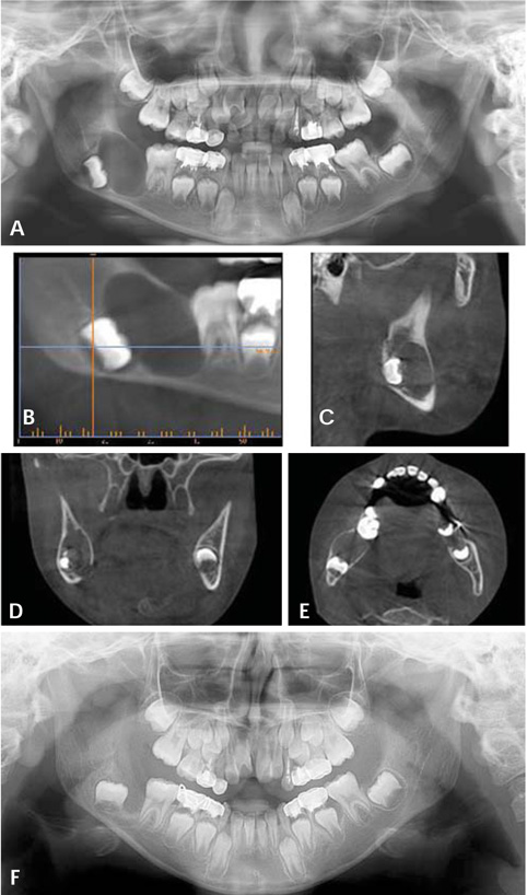
Fig. 1 A. A panoramic radiograph reveals a well-defined round unilocular radiolucency touching the developing crown of the lower right second molar, which is displaced downwardly. The cemento-enamel junction of the involved tooth is incomplete. B-E. The CBCT images show bucco-lingually expanded cortical plates associated with the lesion. F. The 6-month follow-up panoramic radiograph shows uneventful healing of the lesion with upward movement of the more developed lower right second molar.
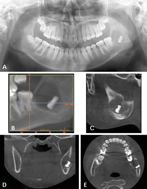
Fig. 2 A. A panoramic radiograph reveals a well-defined round unilocular radiolucency surrounding the developing crown of the lower left third molar. B-E. CBCT images show a lingually expanded cortical plate associated with the lesion, and the buccal cortical plate is perforated.
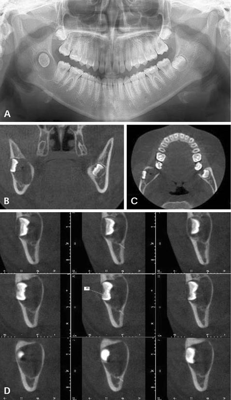
Fig. 3 A. A panoramic radiograph reveals a well-defined round unilocular radiolucency surrounding the transversely positioned crown of the lower right third molar. B-D. CBCT images show a bucco-lingually expanded cystic lesion associated with a buccally displaced developing lower right third molar, but the cementoenamel junction of the crown is incomplete. The mandibular canal is displaced downward due to the lesion.

Fig. 4 A. A panoramic radiograph reveals a large well-defined round unilocular radiolucency surrounding the developing crown of the lower left third molar, which is displaced upward to the sigmoid notch. The cemento-enamel junction of the involved tooth is incomplete. B-C. CBCT images show a bucco-lingually expanded cortical plate associated with the lesion, which surrounds the underdeveloped crown of the lower left third molar.
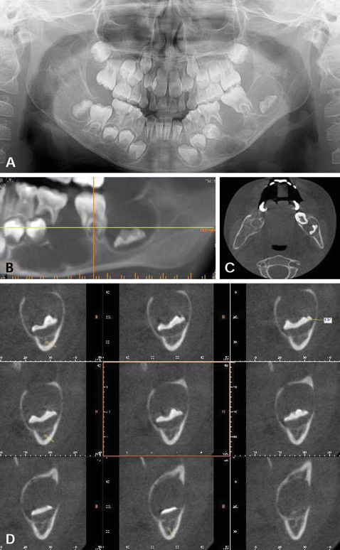
Fig. 5 A. A panoramic radiograph reveals bilateral multiple radiolucencies of the mandible and another separate well-defined round radiolucency involving the developing crown of the lower left second molar. B-D. The CBCT images show bucco-lingually expanded cortical plates associated with the lesion and the mandibular canal is displaced inferiorly and lingually.
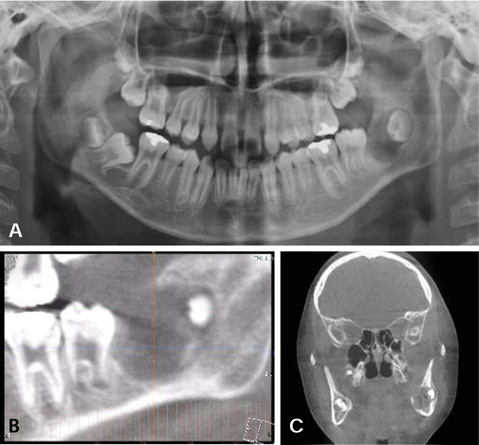
Fig. 6 A. a panoramic radiograph reveals a well-defined round unilocular radiolucency surrounding the developing crown of the lower left third molar, which is displaced distally to the mandibular ramus. The cemento-enamel junction of the tooth is incomplete. B-C. CBCT images show a bucco-lingually expanded cortical plate associated with the lesion, which surrounds the underdeveloped crown of the lower left third molar.

Fig. 7 A. A panoramic radiograph reveals a well-defined round unilocular radiolucency surrounding the developing crown of the lower left third molar without completion of the cemento-enamel junction. B-C. CBCT images show a well-defined round radiolucency with no cortical expansion surrounding the underdeveloped crown of the lower left third molar.

Fig. 8 (A) A panoramic radiograph reveals a well-defined round unilocular radiolucency surrounding the crown of the lower left third molar before the completion of the cemento-enamel junction. (B-D) The CBCT images show a well-defined round radiolucency with no cortical expansion surrounding the underdeveloped crown of the lower left third molar. The mandibular canal can be identified inferior and lingual to the lesion.
Reference
-
1. Neville BW, Damm DD, Allen CM, Bouquot JE. Oral and maxillofacial pathology. 2nd ed. Philadelphia: WB Saunders;2002. p. 590.2. Saluja JS, Ramakrishnan MJ, Vinit GB, Jaiswara C. Multiple dentigerous cysts in a nonsyndromic minor patient: report of an unusual case. Natl J Maxillofac Surg. 2010; 1:168–172.
Article3. Tamgadge A, Tamgadge S, Bhatt D, Bhalerao S, Pereira T, Padhye M. Bilateral dentigerous cyst in a non-syndromic patient: report of an unusual case with review of the literature. J Oral Maxillofac Pathol. 2011; 15:91–95.
Article4. Tsukamoto G, Sasaki A, Akiyama T, Ishikawa T, Kishimoto K, Nishiyama A, et al. A radiologic analysis of dentigerous cysts and odontogenic keratocysts associated with a mandibular third molar. Oral Surg Oral Med Oral Pathol Oral Radiol Endod. 2001; 91:743–747.
Article5. Farah CS, Savage NW. Pericoronal radiolucencies and the significance of early detection. Aust Dent J. 2002; 47:262–265.
Article6. Darling MR, Wehrli BM, Ciavarro C, Daley TD. Pericoronal radiolucency in the posterior mandible. Oral Surg Oral Med Oral Pathol Oral Radiol Endod. 2008; 105:139–143.
Article7. Neville BW, Damm DD, Allen CM, Bouquot JE. Oral and maxillofacial pathology. 2nd ed. Philadelphia: WB Saunders;2002. p. 594–595.8. Curran AE, Damm DD, Drummond JF. Pathologically significant pericoronal lesions in adults: histopathologic evaluation. J Oral Maxillofac Surg. 2002; 60:613–618.
Article9. Vaid N, Kothadiya A, Adwani S. Tooth in a cyst - Is it always a dentigerous cyst? Indian J Otolaryngol Head Neck Surg. 2007; 59:399–400.
Article10. Hemavathy S, Roy S. Follicular odontogenic keratocyst mimicking dentigerous cyst - report of two cases. Arch Oral Sci Res. 2011; 1:100–103.11. Wright BA, Fanibunda K. Odontogenic keratocyst dentigerous cyst type? J Can Dent Assoc. 1981; 47:313–314.12. Yadav S, Verma A, Sheorain A, Sharma A. An unusual case presentation of follicular odontogenic keratocyst with an impacted mesiodens. J Craniofac Surg. 2013; 24:e300–e302.
Article
- Full Text Links
-
- Actions
-
Cited
- CITED
-
- Close
- Share
-
- Similar articles
-
- Several Methods Of Fabrications Of Inner Crown And Outer Crown In Construction Of Konus Denture
- A morphometric study of teeth on the Korean normal occlusion
- Relationship between Pre-Eruptive Buccal Pit Radiolucency and Restoration in Mandibular First Molar
- Incomplete bone formation after sinus augmentation: A case report on radiological findings by computerized tomography at follow-up
- A CLINICAL STUDY OF ODONTECTOMY IN IMPACTED MANDIBULAR THIRD MOLARS
