J Korean Orthop Assoc.
2009 Feb;44(1):123-129. 10.4055/jkoa.2009.44.1.123.
The Surgical Treatment of the Cervical Myelopathy with Laminectomy and Posterior Fusion by using Lateral Mass Screw Fixation
- Affiliations
-
- 1Department of Orthopaedic Surgery, Kyung Hee University College of Medicine, Seoul, Korea. sks111@khmc.or.kr
- 2Department of Orthopaedic Surgery, Seoul Medical Center, Seoul, Korea.
- KMID: 2186387
- DOI: http://doi.org/10.4055/jkoa.2009.44.1.123
Abstract
- PURPOSE
This prospective study was designed to investigate the outcomes of laminectomy and fusion with using lateral mass screw (LMS) fixation for the treatment of cervical myeolpathy.
MATERIALS AND METHODS
We studied a series of 26 consecutive patients with cervical myelopathy and who were planned to undergo laminectomy and fusion with using LMS fixation. MRI was done to investigate the high signal intensity lesion (HSIL) in the cord on the T2 weighted sagittal images. The JOA score, the grip and release test, the finger escape sign, and Hoffman's sign were checked. We analyzed the clinical outcomes depending on the high signal intensity lesion in the cord, the preoperative kyphosis, and the preoperative instability.
RESULTS
The indications for lateral mass screw fixation was kyphotic deformity, segmental instability or ossification of the ligament flavum (OLF). Patients with HSIL on the T2-weighted sagittal MRI was found in 17 patients. The JOA score, the grip and release test, and the finger escape sign were significantly improved after the operation and at the 2 year follow up. The patients with HSIL on the T2-weighted sagittal MRI or segmental instability had a significantly lower preoperative JOA score and a poor postoperative recovery as assessed by the JOA score.
CONCLUSION
Laminectomy and fusion using lateral mass screw fixation for the surgical treatment of cervical myelopathy, which is associated with kyphotic deformity, instability or OLF, is considered a safe and effective treatment option to prevent postoperative kyphosis.
MeSH Terms
Figure
-

Fig. 1 A 60-year-old man who had cervical myelopathy. Preoperative cervical spine lateral radiographs show 5.4° hypolordosis in neutral postion (A), 22.5° kyphosis in flexion (B), and 8.9° lordosis in extension (C). Preoperative MR image (D). Postoperative and 2-year follow up cervical spine lateral radiograph shows recovery of lordosis.
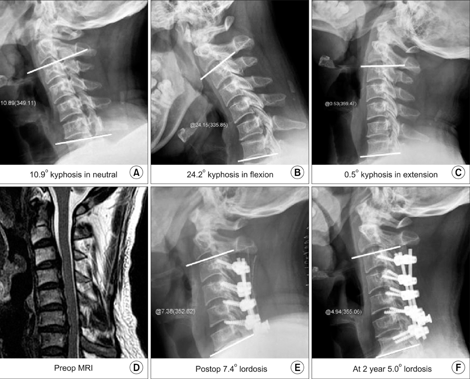
Fig. 2 A 59-year-old man who had cervical myelopathy caused by cervical spondylotic myelopathy with kyphosis. Preoperative cervical spine lateral radiograph shows 10.9° kyphosis in neutral position (A), 24.2° kyphosis in flexion (B), and 0.5° kyphosis in extension (C). Preoperative MR image (D). Postoperative and 2-year follow up cervical spine lateral radiograph shows recovery of lordosis.
Reference
-
1. Iwasaki M, Kawaguchi Y, Kimura T, Yonenobu K. Long-term results of expansive laminoplasty for ossification of the posterior longitudinal ligament of the cervical spine: more than 10 years follow up. J Neurosurg. 2002. 96:Suppl 2. S180–S189.
Article2. Kawaguchi Y, Kanamori M, Ishihara H, Ohmori K, Nakamura H, Kimura T. Minimum 10-year follow up after en bloc cervical laminoplasty. Clin Orthop Relat Res. 2003. 411:129–139.3. Matsunaga S, Sakou T, Nakanisi K. Analysis of the cervical spine alignment following laminoplasty and laminectomy. Spinal Cord. 1999. 37:20–24.
Article4. Muffoletto AJ, Yang J, Vadhva M, Hadjipavlou AG. Cervical stability with lateral mass plating: unicortical versus bicortical screw purchase. Spine. 2003. 28:778–781.5. Ratliff JK, Cooper PR. Cervical laminoplasty: a critical review. J Neurosurg. 2003. 98:230–238.
Article6. Satomi K, Ogawa J, Ishii Y, Hirabayashi K. Short-term complications and lon-term results of expansive open-door laminoplasty for cervical stenotic myelopathy. Spine J. 2001. 1:26–30.7. Shimamura T, Kato S, Toba T, Yamazaki K, Ehara S. Sagittal splitting laminoplasty for spinal canal enlargement for ossification of the spinal ligaments (OPLL and OLF). Semin Musculoskelet Radiol. 2001. 5:203–206.
Article8. Suda K, Abumi K, Ito M, Shono Y, Kaneda K, Fujiya M. Local kyphosis reduces surgical outcomes of expansive open-door laminoplasty for cervical spondylotic myelopathy. Spine. 2003. 28:1258–1262.
Article9. Suda Y, Saitou M, Shioda M, Kohno H, Shibasaki K. Cervical laminoplasty for subaxial lesion in rheumatoid arthritis. J Spinal Disord Tech. 2004. 17:94–101.
Article10. Suk KS, Kim KT, Lee JH, Lee SH, Lim YJ, Kim JS. Sagittal alignment of the cervical spine after the laminoplaty. Spine. 2007. 32:E656–E660.11. Suk KS, Kim KT, Lee SH, Ryu KN. Measurements of lateral mass of cervical spine using MRI for lateral mass screw fixation. J Korean Soc Spine Surg. 2002. 9:121–126.
Article12. Yonenobu K, Hosono N, Iwasaki M, Asano M, Ono K. Neurologic complications of surgery for cervical compression myelopathy. Spine. 1991. 16:1277–1282.
Article
- Full Text Links
-
- Actions
-
Cited
- CITED
-
- Close
- Share
-
- Similar articles
-
- Unusual Anterior Arch Fracture of C1
- C1/2 Transarticular Screw Fixation for Complicated Os Odontoideum: Case Report
- Cervical Myelopathy due to Ossification of Yellow Ligament in a Patient with Reiter's Syndrome
- Posterior Atalntoaxial Fusion with C1 Lateral Mass Screw and C2 Pedicle Screw Supplemented with Miniplate Fixation for Interlaminar Fusion : A Preliminary Report
- Direct Reduction and Fixation for Unstable Hangman's Fracture with Posterior Lateral Mass Plating: Report of 4 Cases
