A Comparative Study of the Navigated and Radiographic Measurements in Open and Closed Wedge High Tibial Osteotomy with Computer Assisted Surgery
- Affiliations
-
- 1Department of Orthopedic Surgery, School of Medicine, Kyung Hee University, Seoul, Korea. songsjun@khmc.or.kr
- KMID: 2186308
- DOI: http://doi.org/10.4055/jkoa.2009.44.5.499
Abstract
- PURPOSE
We wanted to identify the difference of the measured values between a navigation system and radiographs when performing open and closed wedge high tibial osteotomy (HTO) under the control of a navigation system. MATERIALS AND METHODS: Thirty-two open wedge HTOs and 51 closed wedge HTOs were performed using a navigation system. The postoperative mechanical axis percent, which was planned on the navigation system, was 62%. The mechanical axis (MA) was measured before osteotomy and after fixation on the navigation system, and these were compared with the measured values from the radiographs. The difference of the postoperative MA between the navigation system and the radiographs was compared according to the type of HTO. The alteration of the tibial posterior slope angle was also compared. RESULTS: For the open wedge HTO, the mean MA after fixation was valgus 2.7degrees on the navigation system and the postoperative MA was valgus 4.0degrees on the radiograph. For the closed wedge HTO, the mean MA after fixation was valgus 3.5degrees on the navigation system and the postoperative MA was valgus 1.6degrees on the radiograph (p=0.000). The mean tibial posterior slope angle was increased by 5.3degrees after the open wedge HTO and it was decreased by 1.8degrees after closed wedge HTO (p=0.000). CONCLUSION: Performing HTO with a navigation system could increase the surgical accuracy because the navigation system checked the intraoperative correction angle in real time. Weight bearing makes a difference for the postoperative MA between the navigation system and radiographs. This should be taken into account, according to the type of HTO.
MeSH Terms
Figure
-
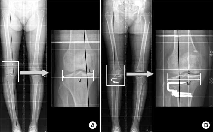
Fig. 1 (A) The mechanical axis % (MA%) shown on the preoperative ortho-roentgenogram is evaluated by percentile denotation [(b/a)×100]. "a" is the width of tibia plateau and "b" is the distance from the medial border of the medial tibial condyle to the point at which the mechanical axis intersects the knee joint line. It shows medial deviation of the mechanical axis. (B) The mechanical axis % (MA%) shown on the postoperative ortho-roentgenogram is evaluated by percentile denotation [(b'/a')×100]. "a'" is the width of tibia plateau and "b'" is the distance from the medial border of the medial tibial condyle to the point at which the mechanical axis intersects the knee joint line. It shows lateral deviation of the mechanical axis.
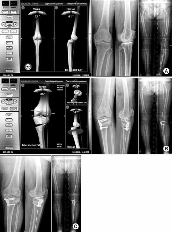
Fig. 2 Change of mechanical axis after the open wedge high tibial osteotomy using navigation system. (A) A 64-year-old woman have the open wedge high tibial osteotomy under navigation control. In the preoperative roentgenogram, the mechanical axis (MA) is varus 9.4° and the mechanical axis % (MA%) is 9.2%. The posterior slope angle of tibia is 8.0°. In the navigation system, the MA is varus 7.4° and the MA% is 22%. (B) In the navigation system, the post-osteotomy MA is valgus 3.5° and the MA% is 57.7%. In the postoperative 2 week roentgenogram, the MA is valgus 5.2° and the MA% is 72.9%. The posterior slope angle of tibia is 12.1°. The postoperative MA and MA% in roentgenogram is larger than the postosteotomy MA and MA% in navigation system. (C) In the postoperative 4 month roentgenogram, the MA is varus 1.2° and the MA% is 37.4%. There are two proximal screws breakage of Puddu plate and loss of correction angle.
Reference
-
1. Coventry MB, Ilstrup DM, Wallrichs SL. Proximal tibial osteotomy. A critical long-term study of eighty-seven cases. J Bone Joint Surg Am. 1993. 75:196–201.
Article2. Giffin JR, Vogrin TM, Zantop T, Woo SL, Harner CD. Effects of increasing tibial slope on the biomechanics of the knee. Am J Sports Med. 2004. 32:376–382.
Article3. Hohmann E, Bryant A, Imhoff AB. The effect of closed wedge high tibial osteotomy on tibial slope: a radiographic study. Knee Surg Sports Traumatol Arthrosc. 2006. 14:454–459.
Article4. Kazakos KJ, Chatzipapas C, Verettas D, Galanis V, Xarchas KC, Psillakis I. Mid-term results of total knee arthroplasty after high tibial osteotomy. Arch Orthop Trauma Surg. 2008. 128:167–173.
Article5. Lee JY, Seon JK, Song EK, Yoon TR, Cheon SY, Lim KY. Comparison of high tibial osteotomy: Opening versus closing wedge osteotomy. J Korean Orthop Assoc. 2004. 39:790–796.
Article6. Nakamura E, Mizuta H, Kudo S, Takagi K, Sakamoto K. Open-wedge osteotomy of the proximal tibia hemicallotasis. J Bone Joint Surg Br. 2001. 83:1111–1115.7. Naudie DD, Amendola A, Fowler PJ. Opening wedge high tibial osteotomy for symptomatic hyperextension-varus thrust. Am J Sports Med. 2004. 32:60–70.
Article8. Brouwer RW, Bierma-Zeinstra SM, van Raaij TM, Verhaar JA. Osteotomy for medial compartment arthritis of the knee using a closing wedge or an opening wedge controlled by a Puddu plate. A one-year randomised controlled study. J Bone Joint Surg Br. 2006. 88:1454–1459.9. Dahl MT. Preoperative planning in deformity correction and limb lengthening surgery. Instr Course Lect. 2000. 49:503–509.10. Ellis RE, Tso CY, Rudan JF, Harrison MM. A surgical planning and guidance system for high tibial osteotomy. Comput Aided Surg. 1999. 4:264–274.
Article11. Hanssen AD. Insall JN, Scott WN, editors. Osteotomy about the knee. Surgery of the knee. 2001. 3rd ed. New York: Churchill Livingstone;1447–1464.12. Kawakami H, Sugano N, Yonenobu K, et al. Effects of rotation on measurement of lower limb alignment for knee osteotomy. J Orthop Res. 2004. 22:1248–1253.
Article13. Keppler P, Gebhard F, Grützner PA, et al. Computer aided high tibial open wedge osteotomy. Injury. 2004. 35:Suppl 1. 68–78.
Article14. Marti CB, Gautier E, Wachtl SW, Jakob RP. Accuracy of frontal and sagittal plane correction in open-wedge high tibial osteotomy. Arthroscopy. 2004. 20:366–372.
Article15. Moreland JR, Bassett LW, Hanker GJ. Radiographic analysis of the axial alignment of the lower extremity. J Bone Joint Surg Am. 1987. 69:745–749.
Article16. Hankemeier S, Hufner T, Wang G, et al. Navigated open-wedge high tibial osteotomy: advantages and disadvantages compared to the conventional technique in a cadaver study. Knee Surg Sports Traumatol Arthrosc. 2006. 14:917–921.
Article17. Wang G, Zheng G, Keppler P, et al. Implementation, accuracy evaluation, and preliminary clinical trial of a CT-free navigation system for high tibial opening wedge osteotomy. Comput Aided Surg. 2005. 10:73–85.
Article18. Bäthis H, Perlick L, Tingart M, Lüring C, Perlick C, Grifka J. Flexion gap configuration in total knee arthroplasty following high tibial osteotomy. Int Orthop. 2004. 28:366–369.19. Puddu G, Franco V. Femoral antivalgus opening wedge osteotomy. Oper Tech Sport Med. 2000. 8:56–60.
Article20. Bae DK, Mun MS, Kwon OS. A newly designed miniplate staple for high tibial osteotomy. Bull Hosp Jt Dis. 1997. 56:167–170.21. Wright JG, Treble N, Feinstein AR. Measurement of lower limb alignment using long radiographs. J Bone Joint Surg Br. 1991. 73:721–723.
Article22. Oswald MH, Jakob RP, Schneider E, Hoogewoud HM. Radiological analysis of normal axial alignment of femur and tibia in view of total knee arthroplasty. J Arthroplasty. 1993. 8:419–426.
Article23. Saragaglia D, Roberts J. Navigated osteotomies around the knee in 170 patients with osteoarthritis secondary to genu varum. Orthopedics. 2005. 28:Suppl. 1269–1274.
Article24. Shaw JA, Dungy DS, Arsht SS. Recurrent varus angulation after high tibial osteotomy: an anatomic analysis. Clin Orthop Relat Res. 2004. 420:205–212.
Article25. Kendoff D, Citak M, Pearle A, et al. Influence of lower limb rotation in navigated alignment anaylsis: implications for high tibial osteotomies. Knee Surg Sports Traumatol Arthrosc. 2007. 15:1003–1008.26. Rodner CM, Adams DJ, Diaz-Doran V, et al. Medial opening wedge tibial osteotomy and the sagittal plane: the effect of increasing tibial slope on tibiofemoral contact pressure. Am J Sports Med. 2006. 34:1431–1441.27. Noyes FR, Goebel SX, West J. Opening wedge tibial osteotomy: the 3-triangle method to correct axial alignment and tibial slope. Am J Sports Med. 2005. 33:378–387.
Article
- Full Text Links
-
- Actions
-
Cited
- CITED
-
- Close
- Share
-
- Similar articles
-
- Navigation versus Radiographic Measurements in the Open-Wedge High Tibial Osteotomy using Computer Assisted Surgery (CAS)
- Comparison of Mechanical Axis and Dynamic Range Assessed with Weight Bearing Radiographs and Navigation System in Closed Wedge High Tibial Osteotomy
- Computer-Assisted Navigation in High Tibial Osteotomy
- Severe Genu Recurvatum after a Closing-wedge High Tibial Osteotomy: A Case Report
- Open-Wedge and Closed-Wedge High Tibial Osteotomy: Current Concept and Long-Term Results
