Comparison of intraoral scanning and conventional impression techniques using 3-dimensional superimposition
- Affiliations
-
- 1Department of Prosthodontics and Research Institute of Oral Science, College of Dentistry, Gangneung-Wonju National University, Gangneung, Republic of Korea. doctorcj@gwnu.ac.kr
- KMID: 2176621
- DOI: http://doi.org/10.4047/jap.2015.7.6.460
Abstract
- PURPOSE
The aim of this study is to evaluate the appropriate impression technique by analyzing the superimposition of 3D digital model for evaluating accuracy of conventional impression technique and digital impression.
MATERIALS AND METHODS
Twenty-four patients who had no periodontitis or temporomandibular joint disease were selected for analysis. As a reference model, digital impressions with a digital impression system were performed. As a test models, for conventional impression dual-arch and full-arch, impression techniques utilizing addition type polyvinylsiloxane for fabrication of cast were applied. 3D laser scanner is used for scanning the cast. Each 3 pairs for 25 STL datasets were imported into the inspection software. The three-dimensional differences were illustrated in a color-coded map. For three-dimensional quantitative analysis, 4 specified contact locations(buccal and lingual cusps of second premolar and molar) were established. For twodimensional quantitative analysis, the sectioning from buccal cusp to lingual cusp of second premolar and molar were acquired depending on the tooth axis.
RESULTS
In color-coded map, the biggest difference between intraoral scanning and dual-arch impression was seen (P<.05). In three-dimensional analysis, the biggest difference was seen between intraoral scanning and dual-arch impression and the smallest difference was seen between dual-arch and full-arch impression.
CONCLUSION
The two- and three-dimensional deviations between intraoral scanner and dual-arch impression was bigger than full-arch and dual-arch impression (P<.05). The second premolar showed significantly bigger three-dimensional deviations than the second molar in the three-dimensional deviations (P>.05).
MeSH Terms
Figure
-
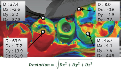
Fig. 1 Three-dimensional divergence(µm) for intraoral scanning (reference) and dual-arch impression (test), divergences in the x-, y-, and z-axes and absolute distance at 4 points.
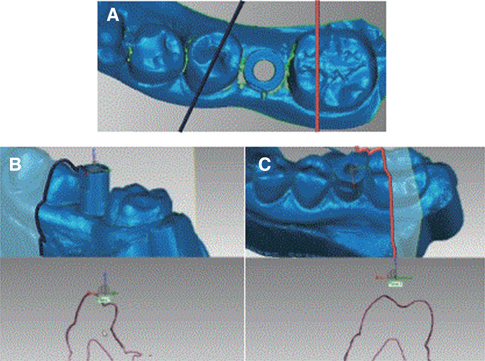
Fig. 2 Two-dimensional divergence for comparing buccolingual and occlusoapical deviations. (A) Reference planes for measurement, (B) Sectional plane for superimposition of second premolars, (C) Sectional plane for superimposition of second molars.
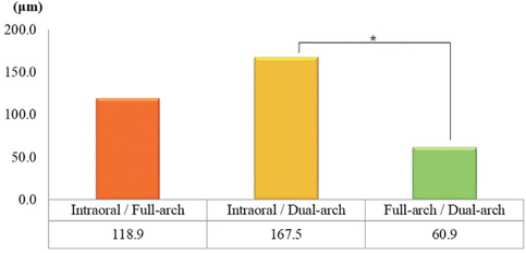
Fig. 3 Comparison of the three-dimensional deviation in second premolar buccal cusp. Asterisks (*) indicates the values that are significantly different among impression groups.
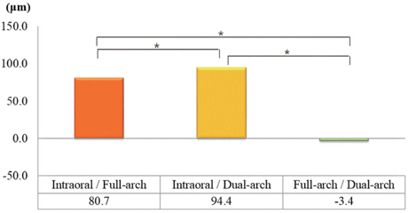
Fig. 4 Comparison of the three-dimensional deviation in second molar buccal cusp. Asterisks (*) indicates the values that are significantly different among impression groups.
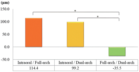
Fig. 5 Comparison of the two-dimensional deviation in second premolar buccal surface. Asterisks (*) indicates the values that are significantly different among impression groups.
Cited by 5 articles
-
Digital evaluation of axial displacement by implant-abutment connection type: An in vitro study
Sung-Jun Kim, KeunBaDa Son, Kyu-Bok Lee
J Adv Prosthodont. 2018;10(5):388-394. doi: 10.4047/jap.2018.10.5.388.Full-arch accuracy of five intraoral scanners:
In vivo analysis of trueness and precision
Miran Kwon, Youngmok Cho, Dong-Wook Kim, MyungSu Kim, Yoon-Ji Kim, Minho Chang
Korean J Orthod. 2021;51(2):95-104. doi: 10.4041/kjod.2021.51.2.95.Accuracy of new implant impression technique using dual arch tray and bite impression coping
Shin-Eon Lee, Sung-Eun Yang, Cheol-Won Lee, Won-Sup Lee, Su Young Lee
J Adv Prosthodont. 2018;10(4):265-270. doi: 10.4047/jap.2018.10.4.265.Verification of a computer-aided replica technique for evaluating prosthesis adaptation using statistical agreement analysis
Hang-Nga Mai, Kyeong Eun Lee, Kyu-Bok Lee, Seung-Mi Jeong, Seok-Jae Lee, Cheong-Hee Lee, Seo-Young An, Du-Hyeong Lee
J Adv Prosthodont. 2017;9(5):358-363. doi: 10.4047/jap.2017.9.5.358.Accuracy evaluation of dental models manufactured by CAD/CAM milling method and 3D printing method
Yoo-Geum Jeong, Wan-Sun Lee, Kyu-Bok Lee
J Adv Prosthodont. 2018;10(3):245-251. doi: 10.4047/jap.2018.10.3.245.
Reference
-
1. The glossary of prosthodontic terms. J Prosthet Dent. 2005; 94:10–92.2. Lee H, So JS, Hochstedler JL, Ercoli C. The accuracy of implant impressions: a systematic review. J Prosthet Dent. 2008; 100:285–291.3. Kan JY, Rungcharassaeng K, Bohsali K, Goodacre CJ, Lang BR. Clinical methods for evaluating implant framework fit. J Prosthet Dent. 1999; 81:7–13.4. Cox JR, Brandt RL, Hughes HJ. The double arch impression technique: a solution to prevent supraocclusion in the indirect restoration. Gen Dent. 2000; 48:86–91.5. Ceyhan JA, Johnson GH, Lepe X. The effect of tray selection, viscosity of impression material, and sequence of pour on the accuracy of dies made from dual-arch impressions. J Prosthet Dent. 2003; 90:143–149.6. Ceyhan JA, Johnson GH, Lepe X, Phillips KM. A clinical study comparing the three-dimensional accuracy of a working die generated from two dual-arch trays and a complete-arch custom tray. J Prosthet Dent. 2003; 90:228–234.7. Hahn SM, Millstein PL, Kinnunen TH, Wright RF. The effect of impression volume and double-arch trays on the registration of maximum intercuspation. J Prosthet Dent. 2009; 102:362–367.8. Kang AH, Johnson GH, Lepe X, Wataha JC. Accuracy of a reformulated fast-set vinyl polysiloxane impression material using dual-arch trays. J Prosthet Dent. 2009; 101:332–341.9. Johnson GH, Mancl LA, Schwedhelm ER, Verhoef DR, Lepe X. Clinical trial investigating success rates for polyether and vinyl polysiloxane impressions made with full-arch and dual-arch plastic trays. J Prosthet Dent. 2010; 103:13–22.10. Parker MH, Cameron SM, Hughbanks JC, Reid DE. Comparison of occlusal contacts in maximum intercuspation for two impression techniques. J Prosthet Dent. 1997; 78:255–259.11. Cox JR, Brandt RL, Hughes HJ. A clinical pilot study of the dimensional accuracy of double-arch and complete-arch impressions. J Prosthet Dent. 2002; 87:510–515.12. Cox JR. A clinical study comparing marginal and occlusal accuracy of crowns fabricated from double-arch and complete-arch impressions. Aust Dent J. 2005; 50:90–94.13. Kaplowitz GJ. Trouble-shooting dual arch impressions II. J Am Dent Assoc. 1997; 128:1277–1281.14. Burke FJ, Crisp RJ. A practice-based assessment of the handling of a fast-setting polyvinyl siloxane impression material used with the dual-arch tray technique. Quintessence Int. 2001; 32:805–810.15. Christensen GJ. Ensuring accuracy and predictability with double-arch impressions. J Am Dent Assoc. 2008; 139:1123–1125.16. Johnson GH, Mancl LA, Schwedhelm ER, Verhoef DR, Lepe X. Clinical trial investigating success rates for polyether and vinyl polysiloxane impressions made with full-arch and dual-arch plastic trays. J Prosthet Dent. 2010; 103:13–22.17. Patzelt SB, Emmanouilidi A, Stampf S, Strub JR, Att W. Accuracy of full-arch scans using intraoral scanners. Clin Oral Investig. 2014; 18:1687–1694.18. Touchstone A, Nieting T, Ulmer N. Digital transition: the collaboration between dentists and laboratory technicians on CAD/CAM restorations. J Am Dent Assoc. 2010; 141:15S–19S.19. Miyazaki T, Hotta Y. CAD/CAM systems available for the fabrication of crown and bridge restorations. Aust Dent J. 2011; 56:97–106.20. Choi HS, Moon JE, Kim SH. The Application of CAD/CAM in Dentistry. J Korean Dent Assoc. 2012; 50:110–117.21. Syrek A, Reich G, Ranftl D, Klein C, Cerny B, Brodesser J. Clinical evaluation of all-ceramic crowns fabricated from intraoral digital impressions based on the principle of active wavefront sampling. J Dent. 2010; 38:553–559.22. Güth JF, Keul C, Stimmelmayr M, Beuer F, Edelhoff D. Accuracy of digital models obtained by direct and indirect data capturing. Clin Oral Investig. 2013; 17:1201–1208.23. Ting-Shu S, Jian S. Intraoral Digital Impression Technique: A Review. J Prosthodont. 2015; 24:313–321.24. Ender A, Mehl A. Accuracy of complete-arch dental impressions: a new method of measuring trueness and precision. J Prosthet Dent. 2013; 109:121–128.25. Seelbach P, Brueckel C, Wöstmann B. Accuracy of digital and conventional impression techniques and workflow. Clin Oral Investig. 2013; 17:1759–1764.26. Quaas S, Rudolph H, Luthardt RG. Direct mechanical data acquisition of dental impressions for the manufacturing of CAD/CAM restorations. J Dent. 2007; 35:903–908.27. da Costa JB, Pelogia F, Hagedorn B, Ferracane JL. Evaluation of different methods of optical impression making on the marginal gap of onlays created with CEREC 3D. Oper Dent. 2010; 35:324–329.28. Christensen GJ. Will digital impressions eliminate the current problems with conventional impressions? J Am Dent Assoc. 2008; 139:761–763.29. Luthardt RG, Kühmstedt P, Walter MH. A new method for the computer-aided evaluation of three-dimensional changes in gypsum materials. Dent Mater. 2003; 19:19–24.30. Price RB, Gerrow JD, Sutow EJ, MacSween R. The dimensional accuracy of 12 impression material and die stone combinations. Int J Prosthodont. 1991; 4:169–174.31. Giménez B, Özcan M, Martínez-Rus F, Pradíes G. Accuracy of a digital impression system based on parallel confocal laser technology for implants with consideration of operator experience and implant angulation and depth. Int J Oral Maxillofac Implants. 2014; 29:853–862.32. Nedelcu RG, Persson AS. Scanning accuracy and precision in 4 intraoral scanners: an in vitro comparison based on 3-dimensional analysis. J Prosthet Dent. 2014; 112:1461–1471.33. Lee SJ, Betensky RA, Gianneschi GE, Gallucci GO. Accuracy of digital versus conventional implant impressions. Clin Oral Implants Res. 2015; 26:715–719.34. Stimmelmayr M, Güth JF, Erdelt K, Edelhoff D, Beuer F. Digital evaluation of the reproducibility of implant scanbody fit--an in vitro study. Clin Oral Investig. 2012; 16:851–856.
- Full Text Links
-
- Actions
-
Cited
- CITED
-
- Close
- Share
-
- Similar articles
-
- Comparison of accuracy between digital and conventional implant impressions: two and three dimensional evaluations
- Comparison of different impression techniques for edentulous jaws using three-dimensional analysis
- Fabricating an Implant-Supported Crown with Impression Scanning Technology
- Implant prosthesis using intraoral scanner: Case Report
- Three dimensional accuracy analysis of dental stone casts fabricated using irreversible hydrocolloid impressions
