A maxillary canine with two separated root canals: a case report
- Affiliations
-
- 1Luden Dental Clinic, Seoul, Korea.
- 2Department of Conservative Dentistry, Kyung Hee University School of Dentistry, Seoul, Korea. shpark94@khu.ac.kr
- 3Department of Conservative Dentistry, Medical Center, Kyung Hee University School of Dentistry, Seoul, Korea.
- 4Department of Endodontics, University of Western Australia, Australia.
- KMID: 2176581
- DOI: http://doi.org/10.5395/JKACD.2011.36.5.431
Abstract
- Maxillary canines have less anatomical diversities than other teeth. They usually have a single root and root canal. This report describes an endodontic treatment of a maxillary canine with two separated root canals which have not been reported through the demonstration of radiography and computerized tomography (CT). Even though appropriated endodontic treatment has been performed, the severe pain could happen due to lack of consideration of anatomical variations of the teeth. Therefore, the clinicians should be well aware of the possibility of anatomical variations in the root canal system during endodontic treatment even if the number of root canals is obvious such as in this case.
Keyword
MeSH Terms
Figure
-
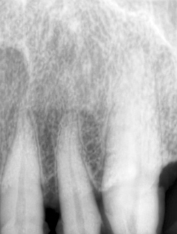
Figure 1 Preoperative radiograph of left maxillary canine. There was no special periapical pathosis.

Figure 2 Saggital plane cone beam CT image of the left maxillary left canine. Additional s-shaped root canal is observed on the palatal side. CT, computed tomograph.

Figure 3 Clinical pictures after access opening. Two separated orifices can be clearly observed.

Figure 4 Files are placed within the root canal to identify the canal path.
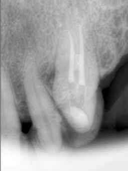
Figure 5 Radiographic image after canal filling. The root canal showed communicating pattern at middle one-third point.
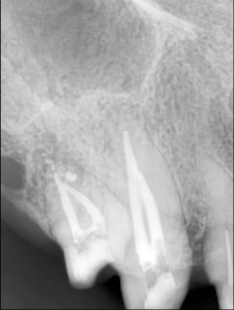
Figure 6 Radiographic image of maxillary right canine of the same patient. Two separated canals converging at the apical one-third point can be seen under the canal filling condition.
Reference
-
1. Sabala CL, Benenati FW, Neas BR. Bilateral root or root canal aberrations in a dental school patient population. J Endod. 1994. 20:38–42.
Article2. Pineda F, Kuttler Y. Mesiodistal and buccolingual roentgenographic investigation of 7,275 root canals. Oral Surg Oral Med Oral Pathol. 1972. 33:101–110.
Article3. Vertucci FJ. Root canal anatomy of the human permanent teeth. Oral Surg Oral Med Oral Pathol. 1984. 58:589–599.
Article4. Calişkan MK, Pehlivan Y, Sepetçioğlu F, Türkün M, Tuncer SS. Root canal morphology of human permanent teeth in a Turkish population. J Endod. 1995. 21:200–204.
Article5. Weng XL, Yu SB, Zhao SL, Wang HG, Mu T, Tang RY, Zhou XD. Root canal morphology of permanent maxillary teeth in the Han nationality in Chinese Guanzhong area: a new modified root canal staining technique. J Endod. 2009. 35:651–656.
Article6. Davis GR, Wong FS. X-ray microtomography of bones and teeth. Physiol Meas. 1996. 17:121–146.
Article7. Alapati S, Zaatar EI, Shyama M, Al-Zuhair N. Maxillary canine with two root canals. Med Princ Pract. 2006. 15:74–76.
Article8. Slowey RR. Radiographic aids in the detection of extra root canals. Oral Surg Oral Med Oral Pathol. 1974. 37:762–772.
Article9. Nair PN, Sjögren U, Krey G, Kahnberg KE, Sundqvist G. Intraradicular bacteria and fungi in root-filled, asymptomatic human teeth with therapy-resistant periapical lesions: a long-term light and electron microscopic follow-up study. J Endod. 1990. 16:580–588.
Article
- Full Text Links
-
- Actions
-
Cited
- CITED
-
- Close
- Share
-
- Similar articles
-
- A Study of Root Canals Morphology in Primary Molars using Computerized Tomography
- Assessment of Root and Root Canal Morphology of Human Primary Molars using CBCT
- Dilemmas pertaining to three canals in the mesiobuccal root of a maxillary second molar: a case report
- Analysis of C-shaped root canal configuration in maxillary molars in a Korean population using cone-beam computed tomography
- Endodontic management of a maxillary first molar with three roots and seven root canals with the aid of cone-beam computed tomography
