Microleakage and characteristics of resin-tooth tissues interface of a selfetch and an etch-and-rinse adhesive systems
- Affiliations
-
- 1Faculty of Odonto-stomatology, University of Medicine and Pharmacy at Ho Chi Minh City (UMP), Ho Chi Minh City, Vietnam
- KMID: 2548074
- DOI: http://doi.org/10.5395/rde.2021.46.e30
Abstract
Objectives
This study was conducted to compare the microleakage and characteristics of the resin-tooth tissue interface between self-etch and etch-and-rinse adhesive systems after 48 hours and 3 months.
Materials and Methods
40 extracted premolar teeth were randomly divided into 2 groups: 1-step self-etch adhesive system – Optibond™ All-In-One, and 2-step etch-and-rinse adhesive system - Adper™ Single Bond 2. Both groups were subjected to 500 thermocycles (5°C–55°C) before scanning electron microscope (SEM) analysis or microleakage trial at 48-hour and 3-month time periods.
Results
SEM images showed the hybrid layer thickness, diameter, and length of resin tags of the self-etch adhesive (0.42 ± 0.14 µm; 1.49 ± 0.45 µm; 16.35 ± 14.26 µm) were smaller than those of the etch-and-rinse adhesive (4.39 ± 1.52 µm; 3.49 ± 1 µm; 52.81 ± 35.81 µm). In dentin, the microleakage scores of the 2 adhesives were not different in both time periods (48 hours/3 months). However, the microleakage score of etch-and-rinse adhesive increased significantly after 3 months (0.8 ± 0.63 and 1.9 ± 0.88, p < 0.05).
Conclusions
The self-etch adhesive exhibited better long-term sealing ability in dentin when compared to that of the etch-and-rinse adhesive. The greater hybrid layer thickness and dimensions of resin tags did not guarantee reliable, long-lasting sealing in the bonding area.
Keyword
Figure
-
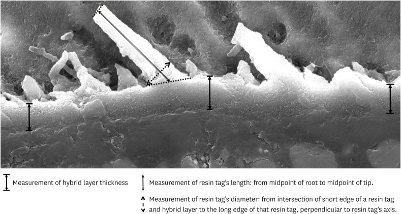
Figure 1 Measurement of hybrid layer, length and diameter of resin tag.
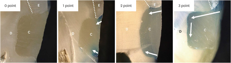
Figure 2 Microleakage score evaluation.C, composite; D, dentin; E, enamel; Arrow, dye penetration.
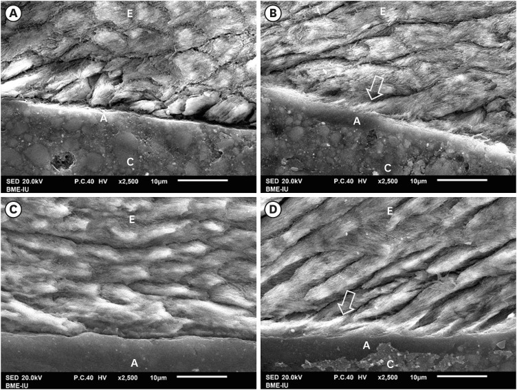
Figure 3 SEM of resin-enamel interface. SEM image of AIO self-etch adhesive after 48 hours (A) and 3 months (C) showing almost flat enamel interface. SB2 etch-and-rinse adhesive after 48 hours (B) and 3 months (D) showing deeply infiltrated resin tags in enamel (arrow).A, adhesive; C, composite; E, enamel.
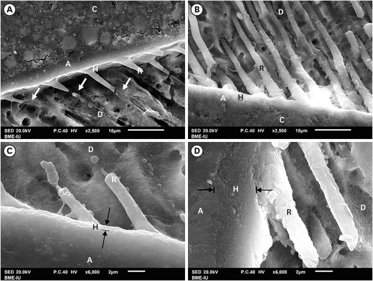
Figure 4 SEM of resin-dentin interface. The AIO self-etch adhesive (A and C) formed thin hybrid layer (between 2 black arrows) (0.42 ± 0.14 µm) with small-diameter (1.49 ± 0.45 µm), short and scattered resin tags (16.35 ± 14.26 µm), some imperfectly formed tags (white arrow). The hybrid layer of the SB2 etch-and-rinse adhesive (B and D) was thick (4.39 ± 1.52 µm) with numerous long (52.81 ± 35.81 µm), large-diameter (3.49 ± 1 µm), funnel-shaped resin tags.A, adhesive; C, composite; D, dentin; H, hybrid layer; R, resin tag.
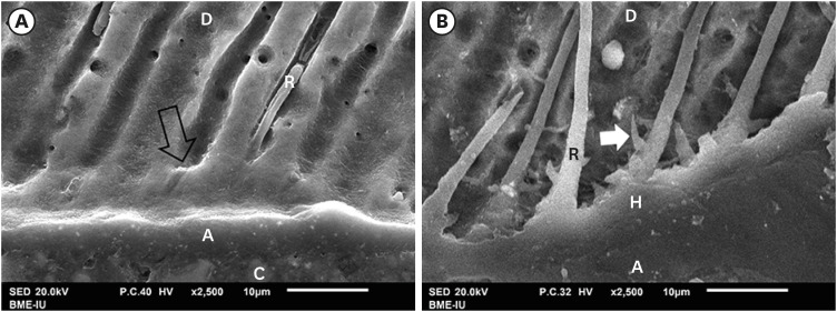
Figure 5 SEM of resin-dentin interface. SEM image of AIO self-etch adhesive (A) showing the “smear plugs” sealing dentin tubules (black arrow) and forming short resin tags. The SB2 etch-and-rinse adhesive (B) showed resin tags infiltrating deeply into dentin tubules and their lateral branches (white arrow).A, adhesive; C, composite; D, dentin; H, hybrid layer; R, resin tag.
Reference
-
1. Buonocore MG. A simple method of increasing the adhesion of acrylic filling materials to enamel surfaces. J Dent Res. 1955; 34:849–853. PMID: 13271655.
Article2. Kugel G, Ferrari M. The science of bonding: from first to sixth generation. J Am Dent Assoc. 2000; 131(Supplement):20S–25S. PMID: 10860341.
Article3. Van Meerbeek B, Yoshihara K, Van Landuyt K, Yoshida Y, Peumans M. From Buonocore's pioneering acid-etch technique to self-adhering restoratives. A Status perspective of rapidly advancing dental adhesive technology. J Adhes Dent. 2020; 22:7–34. PMID: 32030373.4. Sofan E, Sofan A, Palaia G, Tenore G, Romeo U, Migliau G. Classification review of dental adhesive systems: from the IV generation to the universal type. Ann Stomatol (Roma). 2017; 8:1–17. PMID: 28736601.
Article5. Sezinando A. Looking for the ideal adhesive – a review. Rev port Estomatol Med Dent Cir Maxilofac. 2014; 55:194–206.
Article6. Giannini M, Makishi P, Ayres AP, Vermelho PM, Fronza BM, Nikaido T, Tagami J. Self-etch adhesive systems: a literature review. Braz Dent J. 2015; 26:3–10. PMID: 25672377.
Article7. Van Landuyt K, De Munck J, Coutinho E, Peumans M, Lambrechts P, Van Meerbeek B. Bonding to dentin: smear layer and the process of hybridization. In : Eliades G, Watts D, Eliades T, editors. Dental hard tissues and bonding. Berlin, Heidelberg: Springer;2005. p. 89–122.8. Talungchit S, Jessop JL, Cobb DS, Qian F, Geraldeli S, Pashley DH, Armstrong SR. Ethanol-wet bonding and chlorhexidine improve resin-dentin bond durability: quantitative analysis using raman spectroscopy. J Adhes Dent. 2014; 16:441–450. PMID: 25202747.10. Chen C, Niu LN, Xie H, Zhang ZY, Zhou LQ, Jiao K, Chen JH, Pashley DH, Tay FR. Bonding of universal adhesives to dentine--Old wine in new bottles? J Dent. 2015; 43:525–536. PMID: 25797702.11. Moura SK, Reis A, Pelizzaro A, Dal-Bianco K, Loguercio AD, Arana-Chavez VE, Grande RH. Bond strength and morphology of enamel using self-etching adhesive systems with different acidities. J Appl Oral Sci. 2009; 17:315–325. PMID: 19668991.
Article12. Thanaratikul B, Santiwong B, Harnirattisai C. Self-etch or etch-and-rinse mode did not affect the microshear bond strength of a universal adhesive to primary dentin. Dent Mater J. 2016; 35:174–179. PMID: 27041005.
Article13. Van Meerbeek B, Yoshihara K, Yoshida Y, Mine A, De Munck J, Van Landuyt KL. State of the art of self-etch adhesives. Dent Mater. 2011; 27:17–28. PMID: 21109301.
Article14. Abdalla AI, Davidson CL. Shear bond strength and microleakage of new dentin bonding systems. Am J Dent. 1993; 6:295–298. PMID: 7880479.15. Yap AU, Ho KS, Wong KM. Comparison of marginal sealing ability of new generation bonding systems. J Oral Rehabil. 1998; 25:666–671. PMID: 9758395.
Article16. Schuldt C, Birlbauer S, Pitchika V, Crispin A, Hickel R, Kühnisch J. Shear bond strength and microleakage of a self-etching adhesive for fissure sealing after different types of aging. Dent Mater J. 2016; 35:490–497. PMID: 27252006.
Article17. Mohammad ZK, Luddin N, Masudi SM, Alam MK. Microleakage in class II composite restorations bonded with self-etch, one-step, one-component adhesive system: a CLSM study. Int Med J. 1994; 22:418–421.18. Mauro SJ, Durão VC, Briso AL, Sundefeld ML, Rahal V. Effect of different adhesive systems on microleakage in class II composite resin restorations. Int J Adhes Adhes. 2012; 34:6–10.
Article19. Waldman GL, Vaidyanathan TK, Vaidyanathan J. Microleakage and resin-to-dentin interface morphology of pre-etching versus self-etching adhesive systems. Open Dent J. 2008; 2:120–125. PMID: 19444319.
Article20. Armstrong S, Breschi L, Özcan M, Pfefferkorn F, Ferrari M, Van Meerbeek B. Academy of Dental Materials guidance on in vitro testing of dental composite bonding effectiveness to dentin/enamel using micro-tensile bond strength (μTBS) approach. Dent Mater. 2017; 33:133–143. PMID: 28007396.
Article21. Gale MS, Darvell BW. Thermal cycling procedures for laboratory testing of dental restorations. J Dent. 1999; 27:89–99. PMID: 10071465.
Article22. Gateva N, Kabaktchieva R. Hybrid layer thickness in primary and permanent teeth - a comparison between total etch adhesives. J IMAB. 2012; 18:191–199.
Article23. Pereira CN, Daleprane B, Barbosa PF, Moreira AN, de Magalhães CS. Qualitative evaluation of scanning electron microscopy methods in a study of the resin cement/dentine adhesive interface. Microsc Microanal. 2014; 20:268–275. PMID: 24188716.
Article24. Patel MU, Punia SK, Bhat S, Singh G, Bhargava R, Goyal P, Oza S, Raiyani CM. An in vitro evaluation of microleakage of posterior teeth restored with amalgam, composite and zirconomer - a stereomicroscopic study. J Clin Diagn Res. 2015; 9:ZC65–ZC67.25. El Sayed HY, Abdalla AI, Shalby ME. Marginal microleakage of composite resin restorations bonded by desensitizing one step self etch adhesive. Tanta Dent J. 2014; 11:180–188.
Article26. Van Meerbeek B, Dhem A, Goret-Nicaise M, Braem M, Lambrechts P, VanHerle G. Comparative SEM and TEM examination of the ultrastructure of the resin-dentin interdiffusion zone. J Dent Res. 1993; 72:495–501. PMID: 8380820.
Article27. Wieczkowski G Jr, Yu XY, Davis EL, Joynt RB. Microleakage in various dentin bonding agent/composite resin systems. Oper Dent. 1992; (Supplement 5):62–67.28. Nakabayashi N, Takarada K. Effect of HEMA on bonding to dentin. Dent Mater. 1992; 8:125–130. PMID: 1521692.
Article29. Vichi A, Margvelashvili M, Goracci C, Papacchini F, Ferrari M. Bonding and sealing ability of a new self-adhering flowable composite resin in class I restorations. Clin Oral Investig. 2013; 17:1497–1506.
Article30. Tjäderhane L, Nascimento FD, Breschi L, Mazzoni A, Tersariol IL, Geraldeli S, Tezvergil-Mutluay A, Carrilho M, Carvalho RM, Tay FR, Pashley DH. Strategies to prevent hydrolytic degradation of the hybrid layer - a review. Dent Mater. 2013; 29:999–1011. PMID: 23953737.
Article31. Van Landuyt KL, Snauwaert J, De Munck J, Peumans M, Yoshida Y, Poitevin A, Coutinho E, Suzuki K, Lambrechts P, Van Meerbeek B. Systematic review of the chemical composition of contemporary dental adhesives. Biomaterials. 2007; 28:3757–3785. PMID: 17543382.
Article32. Yoshihara K, Nagaoka N, Hayakawa S, Okihara T, Yoshida Y, Van Meerbeek B. Chemical interaction of glycero-phosphate dimethacrylate (GPDM) with hydroxyapatite and dentin. Dent Mater. 2018; 34:1072–1081. PMID: 29716740.
Article33. Kenshima S, Francci C, Reis A, Loguercio AD, Filho LE. Conditioning effect on dentin, resin tags and hybrid layer of different acidity self-etch adhesives applied to thick and thin smear layer. J Dent. 2006; 34:775–783. PMID: 16621219.
Article34. Dickens SH, Cho BH. Interpretation of bond failure through conversion and residual solvent measurements and Weibull analyses of flexural and microtensile bond strengths of bonding agents. Dent Mater. 2005; 21:354–364. PMID: 15766582.
Article35. Sundfeld RH, Valentino TA, de Alexandre RS, Briso AL, Sundefeld ML. Hybrid layer thickness and resin tag length of a self-etching adhesive bonded to sound dentin. J Dent. 2005; 33:675–681. PMID: 16139699.
Article36. Alcantara-Galeana MdCZ, Contreras-Bulnes R, Rodríguez-Vilchis LE, Espinosa-Pesqueira ME, Barrera-Ortega CC, López-Hurtado IM, Fernández-Bobadilla A. Microhardness, structure, and morphology of primary enamel after phosphoric acid, self-etching adhesive, and Er:YAG laser etching. Int J Opt. 2017; 2017:7634739.
Article37. Wang R, Shi Y, Li T, Pan Y, Cui Y, Xia W. Adhesive interfacial characteristics and the related bonding performance of four self-etching adhesives with different functional monomers applied to dentin. J Dent. 2017; 62:72–80. PMID: 28527812.
Article
- Full Text Links
-
- Actions
-
Cited
- CITED
-
- Close
- Share
-
- Similar articles
-
- Marginal microleakage of cervical composite resin restorations bonded using etch-and-rinse and self-etch adhesives: two dimensional vs. three dimensional methods
- Interface between calcium silicate cement and adhesive systems according to adhesive families and cement maturation
- Effect of various bleaching treatments on shear bond strength of different universal adhesives and application modes
- Effect of different air-drying time on the microleakage of single-step self-etch adhesives
- Micro-CT evaluation of internal adaptation in resin fillings with different dentin adhesives
