Biocompatibility of bioaggregate cement on human pulp and periodontal ligament (PDL) derived cells
- Affiliations
-
- 1Department of Orthodontics, Yonsei University College of Dentistry, Seoul, Korea.
- 2Department of Conservative Dentistry, Yonsei University College of Dentistry, Seoul, Korea. shujungshin@yahoo.com
- KMID: 2176469
- DOI: http://doi.org/10.5395/JKACD.2010.35.6.473
Abstract
OBJECTIVES
This study was performed to investigate the biocompatibility of newly introduced Bioaggregate on human pulp and PDL cells.
MATERIALS AND METHODS
Cells were collected from human pulp and PDL tissue of extracted premolars. Cell culture plate was coated either with Bioaggregate or white MTA, then the same number of cells were poured to cell culture dishes. Cell attachment and growth was examined under a phase microscope after 1,3 and 7 days of seeding. Cell viability was measured and the data was analyzed using Student t-test and one way ANOVA.
RESULTS
Both types of cells used in this study were well attached and grew healthy on Bioaggregate and MTA coated culture dishes. No cell inhibition zone was observed in Bioaggregate group. There was no statistical difference of viable cells between bioaggreagte and MTA groups.
CONCLUSIONS
Bioaggregate appeared to be biocompatible compared with white MTA on human pulp and PDL cells.
MeSH Terms
Figure
-

Figure 1 Photos taken from an optical microscope (×40 magnification). The same number of cells (a, human pulp cells; b, PDL cells) were seeded to MTA or Bioaggregate coated cell culture dishes, then initial attachment was observed using a phase microscope. PDL, periodontal ligament; MTA, mineral trioxide aggregate.
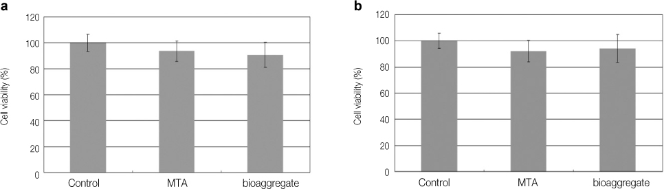
Figure 2 Cell viability tests. a, human pulp cells; b, PDL cells. PDL, periodontal ligament; MTA, mineral trioxide aggregate.
Cited by 2 articles
-
Biocompatibility of root-end filling materials: recent update
Payal Saxena, Saurabh Kumar Gupta, Vilas Newaskar
Restor Dent Endod. 2013;38(3):119-127. doi: 10.5395/rde.2013.38.3.119.Cytotoxicity and physical properties of tricalcium silicate-based endodontic materials
Young-Eun Jang, Bin-Na Lee, Jeong-Tae Koh, Yeong-Joon Park, Nam-Eok Joo, Hoon-Sang Chang, In-Nam Hwang, Won-Mann Oh, Yun-Chan Hwang
Restor Dent Endod. 2014;39(2):89-94. doi: 10.5395/rde.2014.39.2.89.
Reference
-
1. Koh ET, Torabinejad M, Pitt Ford TR, Brady K, McDonald F. Mineral trioxide aggregate stimulates a biological response in human osteoblasts. J Biomed Mater Res. 1997. 37(3):432–439.
Article2. Lee SJ, Monsef M, Torabinejad M. Sealing ability of a mineral trioxide aggregate for repair of lateral root perforations. J Endod. 1993. 19(11):541–544.
Article3. Torabinejad M, Pitt Ford TR, McKendry DJ, Abedi HR, Miller DA, Kariyawasam SP. Histologic assessment of mineral trioxide aggregate as a root-end filling in monkeys. J Endod. 1997. 23(4):225–228.
Article4. Gomes-Filho JE, Rodrigues G, Watanabe S, Estrada Bernabe PF, Lodi CS, Gomes AC, et al. Evaluation of the tissue reaction to fast endodontic cement (CER) and Angelus MTA. J Endod. 2009. 35(10):1377–1380.
Article5. Hashem AA, Hassanien EE. ProRoot MTA, MTA-Angelus and IRM used to repair large furcation perforations: sealability study. J Endod. 2008. 34(1):59–61.
Article6. Song JS, Mante FK, Romanow WJ, Kim S. Chemical analysis of powder and set forms of Portland cement, gray ProRoot MTA, white ProRoot MTA, and gray MTA-Angelus. Oral Surg Oral Med Oral Pathol Oral Radiol Endod. 2006. 102(6):809–815.
Article7. Lessa FC, Aranha AM, Hebling J, Costa CA. Cytotoxic effects of White-MTA and MTA-Bio cements on odontoblast-like cells (MDPC-23). Braz Dent J. 2010. 21(1):24–31.
Article8. Zhang H, Pappen FG, Haapasalo M. Dentin enhances the antibacterial effect of mineral trioxide aggregate and bioaggregate. J Endod. 2009. 35(2):221–224.
Article9. Park JW, Hong SH, Kim JH, Lee SJ, Shin SJ. X-Ray diffraction analysis of white ProRoot MTA and Diadent BioAggregate. Oral Surg Oral Med Oral Pathol Oral Radiol Endod. 2010. 109(1):155–158.
Article10. Vivan RR, Zapata RO, Zeferino MA, Bramante CM, Bernardineli N, Garcia RB, et al. Evaluation of the physical and chemical properties of two commercial and three experimental root-end filling materials. Oral Surg Oral Med Oral Pathol Oral Radiol Endod. 2010. 110(2):250–256.
Article11. Yan P, Yuan Z, Jiang H, Peng B, Bian Z. Effect of bioaggregate on differentiation of human periodontal ligament fibroblasts. Int Endod J. 2010. In press.
Article12. Yuan Z, Peng B, Jiang H, Bian Z, Yan P. Effect of bioaggregate on mineral-associated gene expression in osteoblast cells. J Endod. 2010. 36(7):1145–1148.
Article13. Chang SW, Yoo H, Park D, Oh T, Bae K. Ingredients and cytotoxicity of MTA and 3 kinds of Portland cement. J Korean Acad Conserv Dent. 2008. 33(4):369–376.
Article14. Kwon J, Lim S, Baek S, Bae K, Kang M, Lee W. The effect of mineral trioxide aggregate on the production of growth factors and cytokines by human periondontal ligament fibroblast. J Korean Acad Conserv Dent. 2007. 32(3):191–197.
Article15. Yun YR, Yang I, Hwang Y, Hwang I, Choi H, Yoon S, Kim S, Oh W. Pulp response of mineral trioxide aggregate, calcium sulfate or calcium hydorixde. J Korean Acad Conserv Dent. 2007. 32(2):95–101.
Article16. Apaydin W, Shanahang S, Torabinejad M. Hard-tissue healing after application of fresh or set MTA as root-end filling material. J Endod. 2004. 30(1):21–24.
Article17. Ford T, Torabinejad M, Abedi H, Bakland L, Kariyawasam S. Using mineral trioxide aggregate as a pulp-capping material. J Am Dent Assoc. 1996. 127(10):1491–1494.
Article18. De-Deus G, Canabarro A, Alves G, Linhares A, Senne MI, Granjeiro JM. Optimal cytocompatibility of a bioceramic nanoparticulate cement in primary human mesenchymal cells. J Endod. 2009. 35(10):1387–1390.
Article
- Full Text Links
-
- Actions
-
Cited
- CITED
-
- Close
- Share
-
- Similar articles
-
- A study on differentiation potency of adult stem cells from pulp, periodontal ligament, and dental follicle to osteoblast
- Stem cell properties of cells derived from canine periodontal ligament
- Biochemical characteristics of human periodontal ligament cells in vitro
- Comparison of Gene Expression from Supernumerary Dental Pulp and Periodontal Ligament Stem Cells
- The effects of PDGF and LPS on the viabillty of human periodontal ligament cells
