Cortical and cancellous bone thickness on the anterior region of alveolar bone in Korean: a study of dentate human cadavers
- Affiliations
-
- 1Department of Anatomy and Orofacial Development, School of Dentistry, Chosun University, Gwangju, Korea.
- 2Department of Dental Prosthetics, School of Dentistry, Chosun University, Gwangju, Korea. jhajung@chosun.ac.kr
- KMID: 2176409
- DOI: http://doi.org/10.4047/jap.2012.4.3.146
Abstract
- PURPOSE
The cortical bone thickness on the anterior region is important for achieving implant stability. The purpose of this study was to examine the thickness of the cortical and cancellous bones on the anterior region of the maxilla and mandible.
MATERIALS AND METHODS
Twenty-five cadaver heads were used (16 male and 9 female; mean death age, 56.7 years). After the long axis of alveolar process was set up, it was measured in 5 levels starting from 2 mm below the cementoenamel junction (L1) at intervals of 3 mm. All data was analysed statistically by one-way ANOVA at the .05 significance level.
RESULTS
The cortical bone thickness according to measurement levels in both the labial and lingual sides increased from L1 to L5, and the lingual side below L3 was significantly thicker than the labial side on the maxilla and mandible. In particular, the labial cortical bone thickness in the maxilla was the thinnest compared to the other regions. The cancellous bone thickness according to measurement levels increased from L1 to L5 on the maxilla, and on the mandible it was the thinnest at the middle level of the root.
CONCLUSION
For implant placement on the anterior region, a careful evaluation and full knowledge on the thickness of the cortical and cancellous bone are necessary, therefore, these results may provide an anatomic guideline to clinicians.
Figure
-
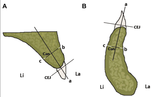
Fig. 1 Diagram showing a cross-section of the interdental area. A: maxilla, B: mandible. a: long axis of alveolar process, b: labial cortical bone thickness, c: lingual cortical bone thickness, Can: cancellous bone thickness, CEJ: cementoenamel junction, La: labial side, Li: lingual side.
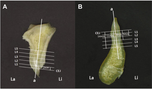
Fig. 2 Scanned image showing the 5 levels starting from 2 mm below the CEJ (L1) at intervals of 3 mm to the root apex area (L5). A: maxilla, B: mandible. a: long axis of alveolar process, CEJ: cementoenamel junction, La: labial side, Li: lingual side. L1: 2 mm below the CEJ, L2: 5 mm below the CEJ, L3: 8 mm below the CEJ, L4: 11 mm below the CEJ, L5: 14 mm below the CEJ.

Fig. 3 Diagram showing that the cortical bone thickness was compared in the labial and lingual sides at each level in the anterior region on the maxilla. *indicates statistical significance with a P<.05.
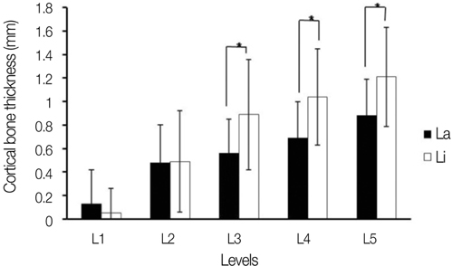
Fig. 4 Diagram showing that the cortical bone thickness was compared in the labial and lingual sides at each level in the anterior region on the mandible. *indicates statistical significance with a P<.05.
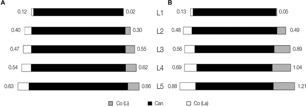
Fig. 5 Diagram showing the average thickness of cortical and cancellous bones at each level in the anterior region. A: maxilla, B: mandible.
Cited by 1 articles
-
Assessment of buccal bone thickness of aesthetic maxillary region: a cone-beam computed tomography study
Ramón Fuentes, Tania Flores, Pablo Navarro, Carlos Salamanca, Víctor Beltrán, Eduardo Borie
J Periodontal Implant Sci. 2015;45(5):162-168. doi: 10.5051/jpis.2015.45.5.162.
Reference
-
1. Ku JE, Yang HS, Yun KD. A morphometric analysis of maxillary central incisor on the basis of facial appearance in Korea. J Adv Prosthodont. 2012. 4:13–17.2. Bernard JP, Schatz JP, Christou P, Belser U, Kiliaridis S. Long-term vertical changes of the anterior maxillary teeth adjacent to single implants in young and mature adults. A retrospective study. J Clin Periodontol. 2004. 31:1024–1028.3. Buser D, Martin W, Belser UC. Optimizing esthetics for implant restorations in the anterior maxilla: anatomic and surgical considerations. Int J Oral Maxillofac Implants. 2004. 19:43–61.4. Miyamoto I, Tsuboi Y, Wada E, Suwa H, Iizuka T. Influence of cortical bone thickness and implant length on implant stability at the time of surgery-clinical, prospective, biomechanical, and imaging study. Bone. 2005. 37:776–780.5. Flanagan D. A comparison of facial and lingual cortical thicknesses in edentulous maxillary and mandibular sites measured on computerized tomograms. J Oral Implantol. 2008. 34:256–258.6. Swasty D, Lee JS, Huang JC, Maki K, Gansky SA, Hatcher D, Miller AJ. Anthropometric analysis of the human mandibular cortical bone as assessed by cone-beam computed tomography. J Oral Maxillofac Surg. 2009. 67:491–500.7. Deguchi T, Nasu M, Murakami K, Yabuuchi T, Kamioka H, Takano-Yamamoto T. Quantitative evaluation of cortical bone thickness with computed tomographic scanning for orthodontic implants. Am J Orthod Dentofacial Orthop. 2006. 129:721.8. Lim WH, Lee SK, Wikesjö UM, Chun YS. A descriptive tissue evaluation at maxillary interradicular sites: implications for orthodontic mini-implant placement. Clin Anat. 2007. 20:760–765.9. Kim JH, Lee JG, Han DH, Kim HJ. Morphometric analysis of the anterior region of the maxillary bone for immediate implant placement using micro-CT. Clin Anat. 2011. 24:462–468.10. Park HD, Min CK, Kwak HH, Youn KH, Choi SH, Kim HJ. Topography of the outer mandibular symphyseal region with reference to the autogenous bone graft. Int J Oral Maxillofac Surg. 2004. 33:781–785.11. Shin JW. Dental anatomy. 2010. 3rd ed. Seoul: DaehanNarae Publishing, Inc.;61–99.12. Katranji A, Misch K, Wang HL. Cortical bone thickness in dentate and edentulous human cadavers. J Periodontol. 2007. 78:874–878.13. Cardaropoli G, Araújo M, Lindhe J. Dynamics of bone tissue formation in tooth extraction sites. An experimental study in dogs. J Clin Periodontol. 2003. 30:809–818.14. Araújo MG, Lindhe J. Dimensional ridge alterations following tooth extraction. An experimental study in the dog. J Clin Periodontol. 2005. 32:212–218.15. Cho YB, Moon SJ, Chung CH, Kim HJ. Resorption of labial bone in maxillary anterior implant. J Adv Prosthodont. 2011. 3:85–89.16. Covani U, Cornelini R, Barone A. Bucco-lingual bone remodeling around implants placed into immediate extraction sockets: a case series. J Periodontol. 2003. 74:268–273.17. Covani U, Cornelini R, Barone A. Vertical crestal bone changes around implants placed into fresh extraction sockets. J Periodontol. 2007. 78:810–815.18. Nevins M, Camelo M, De Paoli S, Friedland B, Schenk RK, Parma-Benfenati S, Simion M, Tinti C, Wagenberg B. A study of the fate of the buccal wall of extraction sockets of teeth with prominent roots. Int J Periodontics Restorative Dent. 2006. 26:19–29.19. Yoo RH, Chuang SK, Erakat MS, Weed M, Dodson TB. Changes in crestal bone levels for immediately loaded implants. Int J Oral Maxillofac Implants. 2006. 21:253–261.20. Covani U, Bortolaia C, Barone A, Sbordone L. Bucco-lingual crestal bone changes after immediate and delayed implant placement. J Periodontol. 2004. 75:1605–1612.21. Araújo MG, Sukekava F, Wennström JL, Lindhe J. Ridge alterations following implant placement in fresh extraction sockets: an experimental study in the dog. J Clin Periodontol. 2005. 32:645–652.22. Araújo MG, Wennström JL, Lindhe J. Modeling of the buccal and lingual bone walls of fresh extraction sites following implant installation. Clin Oral Implants Res. 2006. 17:606–614.23. Cardaropoli G, Lekholm U, Wennström JL. Tissue alterations at implant-supported single-tooth replacements: a 1-year prospective clinical study. Clin Oral Implants Res. 2006. 17:165–171.24. Kan JY, Rungcharassaeng K. Immediate placement and provisionalization of maxillary anterior single implants: a surgical and prosthodontic rationale. Pract Periodontics Aesthet Dent. 2000. 12:817–824.25. Wagenberg BD, Ginsburg TR.. Immediate implant placement on removal of the natural tooth: retrospective analysis of 1,081 implants. Compend Contin Educ Dent. 2001. 22:399–404. 40640826. Juodzbalys G, Wang HL. Soft and hard tissue assessment of immediate implant placement: a case series. Clin Oral Implants Res. 2007. 18:237–243.27. Misch CE. Density of bone: effect on treatment plans, surgical approach, healing, and progressive boen loading. Int J Oral Implantol. 1990. 6:23–31.
- Full Text Links
-
- Actions
-
Cited
- CITED
-
- Close
- Share
-
- Similar articles
-
- Morphometric analysis of maxillary alveolar regions for immediate implantation
- Angulation between Long Axis of Anterior Teeth and Alveolar Process, and Thickness of Alveolar Bone
- Change in bone mineral density according to aging in Korean women: Study of Using Quantitative Computerized Tomography
- A computed tomography-based analysis of the structure of the mandible according to age and sex
- The effects of bone density and crestal cortical bone thickness on micromotion and peri-implant bone strain distribution in an immediately loaded implant: a nonlinear finite element analysis
