J Korean Acad Conserv Dent.
2006 Jan;31(1):50-57. 10.5395/JKACD.2006.31.1.050.
Step by step analysis of root canal instrumentation with ProTaper(R)
- Affiliations
-
- 1Department of Conservative Dentistry, School of Dentistry, Pusan National University, Korea. jeongkil@pusan.ac.kr
- KMID: 2175784
- DOI: http://doi.org/10.5395/JKACD.2006.31.1.050
Abstract
- The purpose of this study was to investigate influence of each file step of ProTaper(R) system on canal transportation. Twenty simulated canals were prepared with either engine-driven ProTaper(R) or manual ProTaper(R). Group R-resin blocks were instrumented with rotary ProTaper(R) and group M-resin blocks were instrumented with manual ProTaper(R). Pre-operative resin blocks and post-operative resin blocks after each file step preparation were scanned. Original canal image and the image after using each file step were superimposed for calculation of centering ratio. The image after using each file step and image after using previous file step were superimposed for calculation of the amount of deviation. Measurements were taken horizontally at five different levels (1, 2, 3, 4 and 5 mm) from the level of apical foramen. In rotary ProTaper(R) instrumentation group, centering ratio and the amount of deviation of each step at all levels were not significantly different (p > 0.05). In manual ProTaper(R) instrumentation group, centering ratio and the amount of deviation of each step at all levels except of 1 mm were not significantly different (p > 0.05). At the level of 1 mm, F2 file step had significantly large centering ratio and the amount of deviation (p < 0.05). Under the condition of this study, F2 file step of manual ProTaper(R) tended to transport the apical part of the canals than that of rotary ProTaper(R).
MeSH Terms
Figure
-
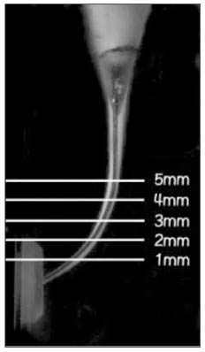
Figure 1 The horizontal lines mean the five measuring levels.

Figure 2 This drawing represents a measuring method. X1 represents the maximum extent of canal movements in one direction and X2 is the movement in the opposite direction. Y is the diameter of prepared canal by each step. Centering ratio = |X1-X2|/Y × 100 Amount of deviation = |X1-X2|
Reference
-
1. European Society of Endodontology. Consensus report of the European Society of Endodontology on quality guidelines for endodontic treatment. Int Endod J. 1994. 27:115–124.2. Schilder H. Cleaning and shaping the root canal. Dent Clin North Am. 1974. 18:269–296.3. Al-Omari MAO, Dummer PMH, Newcombe RG. Comparison of six files to prepare simulated root canals. Part 2. Int Endod J. 1992. 25:67–81.
Article4. Briseño BM, Sonnabend E. The Influence of different root canal instruments on root canal preparation: an in vitro study. Int Endod J. 1991. 24:15–23.
Article5. Schäfer E, Tepal J, Hoppe W. Properties of endodontic hand instruments used in rotary motion. Part 2. Instrumentation of curved canals. J Endod. 1995. 21:493–497.
Article6. Abou-Rass M, Frank AL, Glick DH. The anticurvature filing method to prepare the curved root canal. J Am Dent Assoc. 1980. 101(5):792–794.
Article7. Walia H, Brantley WA, Gerstein H. An initial investigation of the bending and torsional properties of Nitinol root canal files. J Endod. 1988. 14:346–351.
Article8. Camps JJ, Pertot WJ. Tortional and stiffness properties of nickel-titanium K-files. Int Endod J. 1995. 28:239–243.
Article9. Schäfer E. Root canal instruments for manual use: a review. Endod Dent Traumatol. 1997. 13:51–64.
Article10. Glossen CR, Hallor RH, Dove SB, del Rio CE. A comparison of root canal preparations using Ni-Ti hand, Ni-Ti engine driven and K-Flex endodontic instruments. J Endod. 1995. 21:146–151.11. Schäfer E. Shaping ability of Hero 642 rotary Nickel-titanium instruments and stainless steel hand K-Flexofiles in simulated curved root canals. Oral Surg Oral Med Oral Pathol Oral Radiol Endod. 2001. 92(2):215–220.
Article12. Hata G, Uemura M, Kato AS, Imura N, Novo NF, Toda T. A comparison of shaping ability using ProFile, GT file, and Flex-R endodontic instruments in simulated canals. J Endod. 2002. 28(4):316–321.
Article13. Ankrum MT, Hartwell GR, Trutt JE. K3 Endo, ProTaper, and ProFile systems: breakage and distortion in severely curved root of molars. J Endod. 2004. 30(4):234–237.
Article14. Ruddle CJ. The ProTaper Technique: Endodontics Made Easier. Dent Today. 2001. 20(11):58–68.15. Clauder T, Baumann MA. ProTaper NT system. Dent Clin North Am. 2004. 48:87–111.
Article16. Peters OA, Peters CI, Schoneberger K, Barbakov F. ProTaper rotary root canal preparation : effect of canal anatomy on final shape analyzed by micro CT. Int Endod J. 2003. 36:86–92.
Article17. Lee CH, Cho KM, Hong CU. Effect of various canal preparation techniques using rotary Nickel-Titanium files on the maintenance of canal curvature. J Korean Acad Conserv Dent. 2003. 28(1):41–49.
Article18. Schneider SW. A comparison of canal preparations in straight and curved root canals. Oral Surg Oral Med Oral Pathol. 1971. 32(2):271–275.
Article19. Bergmans L, Van Cleynenbreugel J, Beullens M, Wevers M, Van Meerbeek B, Lambrechs P. Progressive versus constant tapered shaft design using NiTi rotary instruments. Int Endod J. 2003. 36:288–295.
Article20. Tulsa Dental Products. ProTaper manufacturer's instructions for use. 2001. Tulsa: Tulsa Dental Products.21. Michael Szasz GDP. ProTaper for hand use-The Experience of a GDP. Dentsply united kingdom - articles. (http://www.dentsply.co.uk).22. Julien Webber. Hand ProTaper-Shaping the Future. Dentsply united kingdom - articles. (http://www.dentsply.co.uk).23. Berutti E, Chiandussi G, Bavaglio I, Ibba A. Comparative analysis of torsional and bending stresses in two mathematical models of nickel-titanium rotary instruments: ProTaper versus ProFile. J Endod. 2003. 29(1):15–19.
Article24. Calberson FL, Deroose CA, Hommez GM, De Moor RJ. Shaping ability of ProTaper nickel-titanium files in simulated resin root canals. Int Endod J. 2004. 37:613–623.
Article25. Iqbal MK, Firic S, Tulcan J, Karabucak B, Kim S. Comparison of apical transportation between ProFile and ProTaper NiTi rotary instruments. Int Endod J. 2004. 37:359–364.
Article26. Yoshimine Y, Ono M, Akamine A. The shaping effects of three nickel-titanium rotary instruments in simulated s-shaped canals. J Endod. 2005. 31(5):373–375.
Article27. Schäfer E, Vlassis M. Comparative investigation of two rotary nickel-titanium instruments: ProTaper versus RaCe. Part 1. Shaping ability in simulated curved canals. Int Endod J. 2004. 37:229–238.
Article28. Yun H, Kim SK. A comparison of the shaping abilities of 4 nickel-titanium rotary instruments in simulated root canals. Oral Surg Oral Med Oral Pathol Oral Radiol Endod. 2003. 95:228–233.
Article
- Full Text Links
-
- Actions
-
Cited
- CITED
-
- Close
- Share
-
- Similar articles
-
- A comparative study on the canal configuration after shaping by ProFile, ProTaper(TM) and K-Flexofile in simulated canals with different angles of curvature
- Comparison of shaping ability using various Nickel-Titanium rotary files and hybrid technique
- Root canal volume change and transportation by Vortex Blue, ProTaper Next, and ProTaper Universal in curved root canals
- Comparison of canal transportation in simulated curved canals prepared with ProTaper Universal and ProTaper Gold systems
- A comparative study of the canal configuration after shaping by protaper rotary and hand files in resin simulated canals
