Root canal volume change and transportation by Vortex Blue, ProTaper Next, and ProTaper Universal in curved root canals
- Affiliations
-
- 1Private Practice, Gyeongju, Korea.
- 2Department of Conservative Dentistry, Wonkwang University Daejeon Dental Hospital, Wonkwang University, Daejeon, Korea. mywizard12@naver.com
- KMID: 2406088
- DOI: http://doi.org/10.5395/rde.2018.43.e3
Abstract
OBJECTIVES
The aim of this study was to compare root canal volume change and canal transportation by Vortex Blue (VB; Dentsply Tulsa Dental Specialties), ProTaper Next (PTN; Dentsply Maillefer), and ProTaper Universal (PTU; Dentsply Maillefer) nickel-titanium rotary files in curved root canals.
MATERIALS AND METHODS
Thirty canals with 20°-45° of curvature from extracted human molars were used. Root canal instrumentation was performed with VB, PTN, and PTU files up to #30.06, X3, and F3, respectively. Changes in root canal volume before and after the instrumentation, and the amount and direction of canal transportation at 1, 3, and 5 mm from the root apex were measured by using micro-computed tomography. Data of canal volume change were statistically analyzed using one-way analysis of variance and Tukey test, while data of amount and direction of transportation were analyzed using Kruskal-Wallis and Mann-Whitney U test.
RESULTS
There were no significant differences among 3 groups in terms of canal volume change (p > 0.05). For the amount of transportation, PTN showed significantly less transportation than PTU at 3 mm level (p = 0.005). VB files showed no significant difference in canal transportation at all 3 levels with either PTN or PTU files. Also, VB files showed unique inward transportation tendency in the apical area.
CONCLUSIONS
Other than PTN produced less amount of transportation than PTU at 3 mm level, all 3 file systems showed similar level of canal volume change and transportation, and VB file system could prepare the curved canals without significant shaping errors.
Keyword
Figure
-
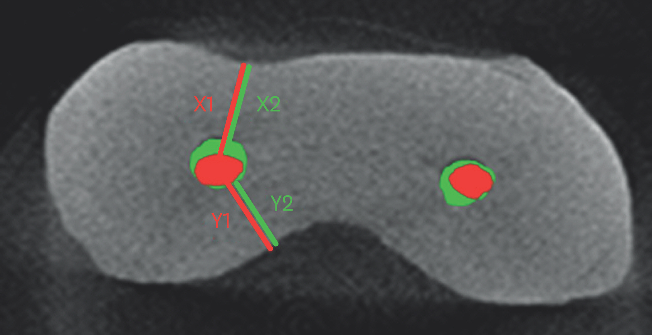
Figure 1. Superimposition of cross-sectional micro-computed tomography (micro-CT) scans before (red) and after (green) the instrumentation for calculating root canal transportation. X is the shortest distance from canal edge to the outside edge of the root, and Y is the shortest distance from canal edge to the inside edge of the root.

Figure 2. Representative 3-dimensional reconstructed micro-computed tomography (micro-CT) images of each group. Before (upper side) and after (lower side) the instrumentation. (A) Vortex Blue, (B) ProTaper Next, and (C) ProTaper Universal.
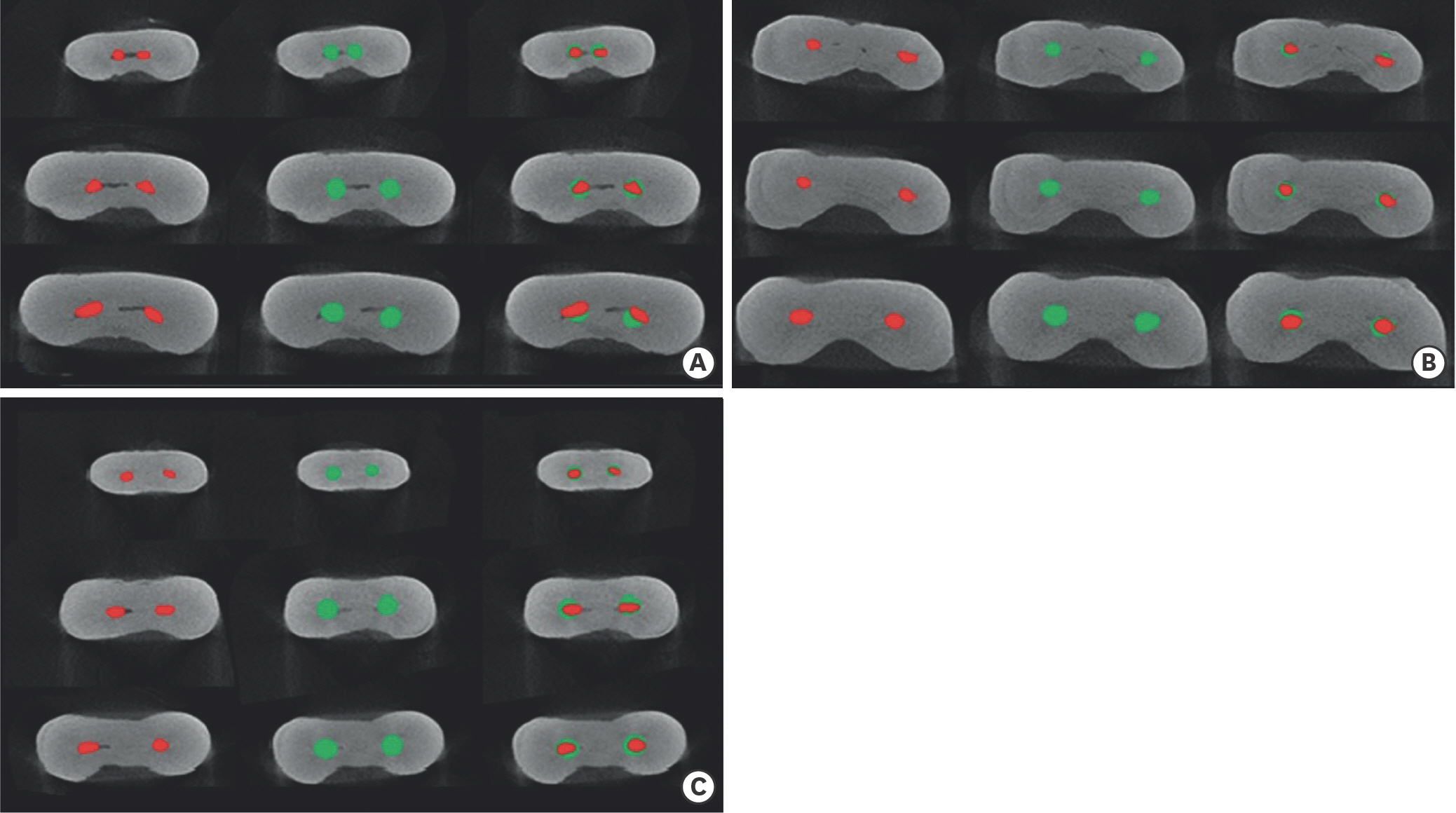
Figure 3. Representative sectional images at the level of 1, 3, and 5 mm from the apex (from top to bottom) of each group. Before (red: left side), after (green: middle side) the instrumentation, and superimposition of the 2 images (right side). (A) Vortex Blue, (B) ProTaper Next, and (C) ProTaper Universal.
Reference
-
References
1. Schäfer E, Bürklein S. Impact of nickel-titanium instrumentation of the root canal on clinical outcomes: a focused review. Odontology. 2012; 100:130–136.
Article2. Elnaghy AM. Cyclic fatigue resistance of ProTaper Next nickel-titanium rotary files. Int Endod J. 2014; 47:1034–1039.
Article3. Kim HC, Sung SY, Ha JH, Solomonov M, Lee JM, Lee CJ, Kim BM. Stress generation during self-adjusting file movement: minimally invasive instrumentation. J Endod. 2013; 39:1572–1575.
Article4. Pirani C, Iacono F, Generali L, Sassatelli P, Nucci C, Lusvarghi L, Gandolfi MG, Prati C. HyFlex EDM: superficial features, metallurgical analysis and fatigue resistance of innovative electro discharge machined NiTi rotary instruments. Int Endod J. 2016; 49:483–493.
Article5. Bonessio N, Pereira ES, Lomiento G, Arias A, Bahia MG, Buono VT, Peters OA. Validated finite element analyses of WaveOne Endodontic Instruments: a comparison between M-Wire and NiTi alloys. Int Endod J. 2015; 48:441–450.
Article6. McRay B, Cox TC, Cohenca N, Johnson JD, Paranjpe A. A microcomputed tomography-based comparison of the canal transportation and centering ability of ProTaper Universal rotary and WaveOne reciprocating files. Quintessence Int. 2014; 45:101–108.7. Nguyen HH, Fong H, Paranjpe A, Flake NM, Johnson JD, Peters OA. Evaluation of the resistance to cyclic fatigue among ProTaper Next, ProTaper Universal, and Vortex Blue rotary instruments. J Endod. 2014; 40:1190–1193.
Article8. Plotino G, Grande NM, Cotti E, Testarelli L, Gambarini G. Blue treatment enhances cyclic fatigue resistance of vortex nickel-titanium rotary files. J Endod. 2014; 40:1451–1453.
Article9. Shen Y, Zhou H, Coil JM, Aljazaeri B, Buttar R, Wang Z, Zheng YF, Haapasalo M. ProFile Vortex and Vortex Blue nickel-titanium rotary instruments after clinical use. J Endod. 2015; 41:937–942.
Article10. Capar ID, Ertas H, Ok E, Arslan H, Ertas ET. Comparative study of different novel nickel-titanium rotary systems for root canal preparation in severely curved root canals. J Endod. 2014; 40:852–856.
Article11. Uygun AD, Kol E, Topcu MK, Seckin F, Ersoy I, Tanriver M. Variations in cyclic fatigue resistance among ProTaper Gold, ProTaper Next and ProTaper Universal instruments at different levels. Int Endod J. 2016; 49:494–499.
Article12. Gagliardi J, Versiani MA, de Sousa-Neto MD, Plazas-Garzon A, Basrani B. Evaluation of the shaping characteristics of ProTaper Gold, ProTaper NEXT, and ProTaper Universal in curved canals. J Endod. 2015; 41:1718–1724.
Article13. Gao Y, Gutmann JL, Wilkinson K, Maxwell R, Ammon D. Evaluation of the impact of raw materials on the fatigue and mechanical properties of ProFile Vortex rotary instruments. J Endod. 2012; 38:398–401.
Article14. Schneider SW. A comparison of canal preparations in straight and curved root canals. Oral Surg Oral Med Oral Pathol. 1971; 32:271–275.
Article15. Gambill JM, Alder M, del Rio CE. Comparison of nickel-titanium and stainless steel hand-file instrumentation using computed tomography. J Endod. 1996; 22:369–375.
Article16. You SY, Kim HC, Bae KS, Baek SH, Kum KY, Lee W. Shaping ability of reciprocating motion in curved root canals: a comparative study with microcomputed tomography. J Endod. 2011; 37:1296–1300.
Article17. Celikten B, Uzuntas CF, Orhan AI, Tufenkci P, Misirli M, Demiralp KO, Orhan K. Micro-CT assessment of the sealing ability of three root canal filling techniques. J Oral Sci. 2015; 57:361–366.
Article18. Marceliano-Alves MF, Sousa-Neto MD, Fidel SR, Steier L, Robinson JP, Pécora JD, Versiani MA. Shaping ability of single-file reciprocating and heat-treated multifile rotary systems: a micro-CT study. Int Endod J. 2015; 48:1129–1136.
Article19. Huang CC, Chang YC, Chuang MC, Lin HJ, Tsai YL, Chang SH, Chen JC, Jeng JH. Analysis of the width of vertical root fracture in endodontically treated teeth by 2 microcomputed tomography systems. J Endod. 2014; 40:698–702.
Article20. Siqueira JF Jr, Alves FR, Almeida BM, de Oliveira JC, Rôças IN. Ability of chemomechanical preparation with either rotary instruments or self-adjusting file to disinfect oval-shaped root canals. J Endod. 2010; 36:1860–1865.
Article21. Tang W, Wu Y, Smales RJ. Identifying and reducing risks for potential fractures in endodontically treated teeth. J Endod. 2010; 36:609–617.
Article22. Pasqualini D, Alovisi M, Cemenasco A, Mancini L, Paolino DS, Bianchi CC, Roggia A, Scotti N, Berutti E. Micro-Computed tomography evaluation of ProTaper Next and BioRace shaping outcomes in maxillary first molar curved canals. J Endod. 2015; 41:1706–1710.
Article23. Zhao D, Shen Y, Peng B, Haapasalo M. Root canal preparation of mandibular molars with 3 nickel-titanium rotary instruments: a microcomputed tomographic study. J Endod. 2014; 40:1860–1864.24. Cruz A, Vera J, Gascón G, Palafox-Sánchez CA, Amezcua O, Mercado G. Debris remaining in the apical third of root canals after chemomechanical preparation by using sodium hypochlorite and glyde: an in vivo study. J Endod. 2014; 40:1419–1423.25. Bürklein S, Mathey D, Schäfer E. Shaping ability of ProTaper NEXT and BT-RaCe nickel-titanium instruments in severely curved root canals. Int Endod J. 2015; 48:774–781.
Article26. Celikten B, Uzuntas CF, Kursun S, Orhan AI, Tufenkci P, Orhan K, Demiralp KO. Comparative evaluation of shaping ability of two nickel-titanium rotary systems using cone beam computed tomography. BMC Oral Health. 2015; 15:32.
Article27. Kandaswamy D, Venkateshbabu N, Porkodi I, Pradeep G. Canal-centering ability: an endodontic challenge. J Conserv Dent. 2009; 12:3–9.
Article28. Alapati SB, Brantley WA, Iijima M, Clark WA, Kovarik L, Buie C, Liu J, Ben Johnson W. Metallurgical characterization of a new nickel-titanium wire for rotary endodontic instruments. J Endod. 2009; 35:1589–1593.
Article29. Shen Y, Zhou HM, Zheng YF, Peng B, Haapasalo M. Current challenges and concepts of the thermomechanical treatment of nickel-titanium instruments. J Endod. 2013; 39:163–172.
Article30. Duke F, Shen Y, Zhou H, Ruse ND, Wang ZJ, Hieawy A, Haapasalo M. Cyclic fatigue of ProFile Vortex and Vortex Blue nickel-titanium files in single and double curvatures. J Endod. 2015; 41:1686–1690.
Article31. Dummer PM, Alodeh MH, al-Omari MA. A method for the construction of simulated root canals in clear resin blocks. Int Endod J. 1991; 24:63–66.
Article32. Lim YJ, Park SJ, Kim HC, Min KS. Comparison of the centering ability of Wave·One and Reciproc nickel-titanium instruments in simulated curved canals. Restor Dent Endod. 2013; 38:21–25.
Article33. Yoo YS, Cho YB. A comparison of the shaping ability of reciprocating NiTi instruments in simulated curved canals. Restor Dent Endod. 2012; 37:220–227.
Article34. Peters OA, Peters CI, Schönenberger K, Barbakow F. ProTaper rotary root canal preparation: effects of canal anatomy on final shape analysed by micro CT. Int Endod J. 2003; 36:86–92.
Article35. Ha JH, Lee CJ, Kwak SW, El Abed R, Ha D, Kim HC. Geometric optimization for development of glide path preparation nickel-titanium rotary instrument. J Endod. 2015; 41:916–919.
Article36. Berutti E, Paolino DS, Chiandussi G, Alovisi M, Cantatore G, Castellucci A, Pasqualini D. Root canal anatomy preservation of WaveOne reciprocating files with or without glide path. J Endod. 2012; 38:101–104.
Article37. Pagliosa A, Sousa-Neto MD, Versiani MA, Raucci-Neto W, Silva-Sousa YT, Alfredo E. Computed tomography evaluation of rotary systems on the root canal transportation and centering ability. Braz Oral Res. 2015; 29:1–7.
Article38. Weine FS, Kelly RF, Lio PJ. The effect of preparation procedures on original canal shape and on apical foramen shape. J Endod. 1975; 1:255–262.
Article39. Wu MK, Fan B, Wesselink PR. Leakage along apical root fillings in curved root canals. Part I: effects of apical transportation on seal of root fillings. J Endod. 2000; 26:210–216.
Article
- Full Text Links
-
- Actions
-
Cited
- CITED
-
- Close
- Share
-
- Similar articles
-
- Comparison of shaping ability using various Nickel-Titanium rotary files and hybrid technique
- Comparison of canal transportation in simulated curved canals prepared with ProTaper Universal and ProTaper Gold systems
- Relative efficacy of three Ni-Ti file systems used by undergraduates
- A comparative study of the canal configuration after shaping by protaper rotary and hand files in resin simulated canals
- Step by step analysis of root canal instrumentation with ProTaper(R)
