Investig Magn Reson Imaging.
2015 Jun;19(2):114-116. 10.13104/imri.2015.19.2.114.
Lack of Myelination in the Anterior Limbs of the Internal Capsule Associated with Cri-du-Chat Syndrome: Case Report
- Affiliations
-
- 1Department of Radiology, Chungnam National University Hospital, Daejeon, Korea. sunkyou@cnuh.co.kr
- 2Department of Radiology, Seoul National University Children's Hospital, Seoul, Korea.
- KMID: 2175591
- DOI: http://doi.org/10.13104/imri.2015.19.2.114
Abstract
- A 21-month-old girl with cri-du-chat syndrome in conjunction with developmental delay underwent brain magnetic resonance imaging (MRI). The MRI showed hypoplasia of the brain stem, a normal cerebellum, thinning of the corpus callosum, and a lack of myelination in both anterior limbs of the internal capsule. She also had neonatal bilateral subependymal cysts. We believe that the symmetrical lack of myelination in both anterior limbs of the internal capsule could be a diagnostic clue of cri-du-chat syndrome.
Keyword
MeSH Terms
Figure
-
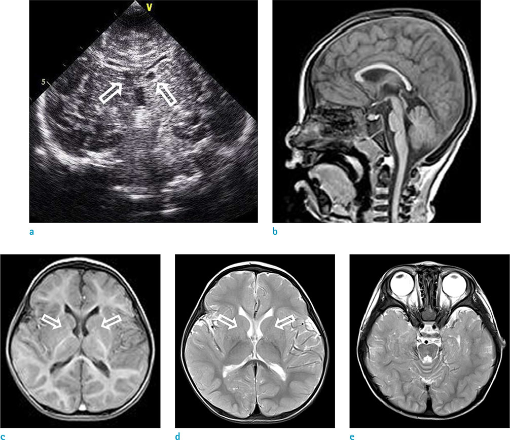
Fig. 1 Neuroradiological findings of a female child with cri-du-chat syndrome. (a) Coronal cranial US on the second day after birth shows subependymal cysts (arrows). Sagittal, axial T1-weighted (b, c) and axial T2-weighted images (d, e) obtained at 21 months showing hypoplasia of the brain stem, a normal cerebellum, thinning of the corpus callosum, and lack of myelination in in both anterior limbs of the internal capsule (arrows).
Reference
-
1. Rodriguez-Caballero A, Torres-Lagares D, Rodriguez-Perez A, Serrera-Figallo MA, Hernandez-Guisado JM, Machuca-Portillo G. Cri du chat syndrome: a critical review. Med Oral Patol Oral Cir Bucal. 2010; 15:e473–e478.2. Lejeune J, Lafourcade J, Berger R, et al. 3 cases of partial deletion of the short arm of a 5 chromosome. C R Hebd Seances Acad Sci. 1963; 257:3098–3102.3. Ninchoji T, Takanashi J. Pontine hypoplasia in 5p-syndrome: a key MRI finding for a diagnosis. Brain Dev. 2010; 32:571–573.4. Uzunhan TA, Sayınbatur B, Calıskan M, Sahin A, Aydın K. A clue in the diagnosis of Cri-du-chat syndrome: Pontine hypoplasia. Ann Indian Acad Neurol. 2014; 17:209–210.5. Hong JH, Lee HY, Lim MK, et al. Brain stem hypoplasia associated with Cri-du-Chat syndrome. Korean J Radiol. 2013; 14:960–962.6. Tamraz J, Rethore M, Lejeune J, et al. Brain morphometry using MRI in cri-du-chat syndrome. Report of seven cases with review of the literature. Ann Genet. 1993; 36:75–87.7. De Michele G, Presta M, Di Salle F, et al. Cerebellar vermis hypoplasia in a case of cri-du-chat syndrome. Acta Neurol (Napoli). 1993; 15:92–96.8. Teoh XH, Tan TY, Chow KK, Lee IW. Prenatal diagnosis of cri-du-chat syndrome: importance of ultrasonographical markers. Singapore Med J. 2009; 50:e181–e184.
