J Gynecol Oncol.
2010 Mar;21(1):18-23. 10.3802/jgo.2010.21.1.18.
Clinical significance of tumor volume and lymph node involvement assessed by MRI in stage IIB cervical cancer patients treated with concurrent chemoradiation therapy
- Affiliations
-
- 1Division of Gynecologic Oncology, Department of Obstetrics and Gynecology, Yonsei University College of Medicine, Seoul, Korea. ytkchoi@yuhs.ac
- 2Division of Gynecologic Oncology, Department of Obstetrics and Gynecology, Kwandong University College of Medicine, Seoul, Korea.
- KMID: 2173505
- DOI: http://doi.org/10.3802/jgo.2010.21.1.18
Abstract
OBJECTIVE
The purpose of this study was to evaluate the prognostic significance of tumor volume assessed by pretreatment MRI in stage IIB cervical cancer patients with concurrent chemoradiation therapy. METHODS: A retrospective chart review was performed on seventy five patients with cervical cancer who were treated with concurrent weekly cisplatin (40 mg/m2) and radiotherapy between January 2000 and April 2007. Potential prognostic factors were age, chemotherapy numbers, histology, tumor diameter and volume, lymph node (LN) involvement and pretreatment squamous cell carcinoma antigen (SCC-Ag) levels. RESULTS: The median follow-up time was 55 months (range, 8 to 104 months). The median tumor size and volume (range) were 4.5 cm (2 to 10) and 33.1 mL (4.2 to 392.7), respectively. Pelvic LN enlargement rate was 58.7%. Para-aortic LN enlargement rate was 14.7%. Using multivariate analysis, a tumor volume (>33 mL, p=0.025), pelvic LN enlargement (p=0.044) revealed a significantly unfavorable outcome on overall survival. PFS was influenced by tumor histology (p<0.001), pelvic LN enlargement (p=0.015) and pretreatment SCC-Ag levels (p=0.018). We found that 22 (29.3%) patients had recurrences and 14 (18.7%) patients died of disease. The 5-year overall survival rate was 80.6% (standard error, 4.9%) and 5-year PFS rate was 71.3% (standard error, 5.3%). CONCLUSION: Tumor volume and pelvic LN involvement showed possibility to predict overall survival in patient with stage IIB cervical cancer. Optimal tumor volume and pelvic LN assessment by pretreatment MRI might be helpful to predict treatment outcome.
Keyword
MeSH Terms
Figure
-
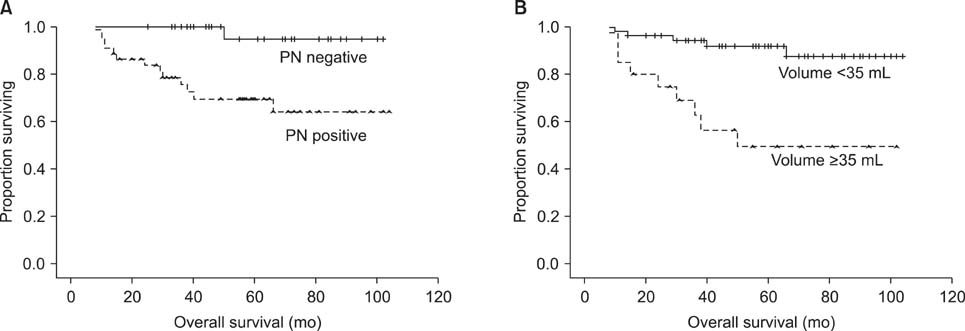
Fig. 1 Overall survival curves for (A) pelvic LN involvement (left, p=0.044) and (B) tumor volume (right, p=0.025). PN: pelvic lymph node.
Reference
-
1. Hricak H, Yu KK. Radiology in invasive cervical cancer. AJR Am J Roentgenol. 1996. 167:1101–1108.2. National Cancer Institute. NCI issues clinical announcement on cervical cancer: chemotherapy plus radiation improves survival. 1999. Bethesda: National Cancer Institute.3. Eifel PJ, Morris M, Wharton JT, Oswald MJ. The influence of tumor size and morphology on the outcome of patients with FIGO stage IB squamous cell carcinoma of the uterine cervix. Int J Radiat Oncol Biol Phys. 1994. 29:9–16.4. Burghardt E, Baltzer J, Tulusan AH, Haas J. Results of surgical treatment of 1028 cervical cancers studied with volumetry. Cancer. 1992. 70:648–655.5. Micke O, Bruns F, Schafer U, Prott FJ, Willich N. The impact of squamous cell carcinoma (SCC) antigen in patients with advanced cancer of uterine cervix treated with (chemo-)radiotherapy. Anticancer Res. 2005. 25:1663–1666.6. Ogino I, Nakayama H, Okamoto N, Kitamura T, Inoue T. The role of pretreatment squamous cell carcinoma antigen level in locally advanced squamous cell carcinoma of the uterine cervix treated by radiotherapy. Int J Gynecol Cancer. 2006. 16:1094–1100.7. Scheidler J, Heuck AF. Imaging of cancer of the cervix. Radiol Clin North Am. 2002. 40:577–590.8. Amendola MA, Hricak H, Mitchell DG, Snyder B, Chi DS, Long HJ 3rd, et al. Utilization of diagnostic studies in the pretreatment evaluation of invasive cervical cancer in the United States: results of intergroup protocol ACRIN 6651/GOG 183. J Clin Oncol. 2005. 23:7454–7459.9. Thoms WW Jr, Eifel PJ, Smith TL, Morris M, Delclos L, Wharton JT, et al. Bulky endocervical carcinoma: a 23-year experience. Int J Radiat Oncol Biol Phys. 1992. 23:491–499.10. Ebner F, Tamussino K, Kressel HY. Magnetic resonance imaging in cervical carcinoma: diagnosis, staging, and follow-up. Magn Reson Q. 1994. 10:22–42.11. Narayan K, McKenzie A, Fisher R, Susil B, Jobling T, Bernshaw D. Estimation of tumor volume in cervical cancer by magnetic resonance imaging. Am J Clin Oncol. 2003. 26:e163–e168.12. Oellinger JJ, Blohmer JU, Michniewicz K, Siewert C, Wust P, Gutberlet M, et al. Pre-operative staging of cervical cancer: comparison of magnetic resonance imaging (MRI) and computed tomography (CT) with histologic results. Zentralbl Gynakol. 2000. 122:82–91.13. Zaino RJ, Ward S, Delgado G, Bundy B, Gore H, Fetter G, et al. Histopathologic predictors of the behavior of surgically treated stage IB squamous cell carcinoma of the cervix: a Gynecologic Oncology Group study. Cancer. 1992. 69:1750–1758.14. Choi SH, Kim SH, Choi HJ, Park BK, Lee HJ. Preoperative magnetic resonance imaging staging of uterine cervical carcinoma: results of prospective study. J Comput Assist Tomogr. 2004. 28:620–627.15. Yu KK, Hricak H, Subak LL, Zaloudek CJ, Powell CB. Preoperative staging of cervical carcinoma: phased array coil fast spin-echo versus body coil spin-echo T2-weighted MR imaging. AJR Am J Roentgenol. 1998. 171:707–711.16. Kawagoe T, Kashimura M, Matsuura Y, Sugihara K, Toki N, Aoki T. Clinical significance of tumor size in stage IB and II carcinoma of the uterine cervix. Int J Gynecol Cancer. 1999. 9:421–426.17. Sakuragi N, Satoh C, Takeda N, Hareyama H, Takeda M, Yamamoto R, et al. Incidence and distribution pattern of pelvic and paraaortic lymph node metastasis in patients with Stages IB, IIA, and IIB cervical carcinoma treated with radical hysterectomy. Cancer. 1999. 85:1547–1554.18. Takeda N, Sakuragi N, Takeda M, Okamoto K, Kuwabara M, Negishi H, et al. Multivariate analysis of histopathologic prognostic factors for invasive cervical cancer treated with radical hysterectomy and systematic retroperitoneal lymphadenectomy. Acta Obstet Gynecol Scand. 2002. 81:1144–1151.19. Tinga DJ, Timmer PR, Bouma J, Aalders JG. Prognostic significance of single versus multiple lymph node metastases in cervical carcinoma stage IB. Gynecol Oncol. 1990. 39:175–180.20. Inoue T, Morita K. The prognostic significance of number of positive nodes in cervical carcinoma stages IB, IIA, and IIB. Cancer. 1990. 65:1923–1927.21. Kamura T, Tsukamoto N, Tsuruchi N, Kaku T, Saito T, To N, et al. Histopathologic prognostic factors in stage IIb cervical carcinoma treated with radical hysterectomy and pelvic-node dissection: an analysis with mathematical statistics. Int J Gynecol Cancer. 1993. 3:219–225.22. Suprasert P, Srisomboon J, Kasamatsu T. Radical hysterectomy for stage IIB cervical cancer: a review. Int J Gynecol Cancer. 2005. 15:995–1001.23. Rose PG, Adler LP, Rodriguez M, Faulhaber PF, Abdul-Karim FW, Miraldi F. Positron emission tomography for evaluating para-aortic nodal metastasis in locally advanced cervical cancer before surgical staging: a surgicopathologic study. J Clin Oncol. 1999. 17:41–45.24. Narayan K, Hicks RJ, Jobling T, Bernshaw D, McKenzie AF. A comparison of MRI and PET scanning in surgically staged loco-regionally advanced cervical cancer: potential impact on treatment. Int J Gynecol Cancer. 2001. 11:263–271.25. Mayr NA, Taoka T, Yuh WT, Denning LM, Zhen WK, Paulino AC, et al. Method and timing of tumor volume measurement for outcome prediction in cervical cancer using magnetic resonance imaging. Int J Radiat Oncol Biol Phys. 2002. 52:14–22.26. Hatano K, Sekiya Y, Araki H, Sakai M, Togawa T, Narita Y, et al. Evaluation of the therapeutic effect of radiotherapy on cervical cancer using magnetic resonance imaging. Int J Radiat Oncol Biol Phys. 1999. 45:639–644.27. Lea JS, Sheets EE, Wenham RM, Duska LR, Coleman RL, Miller DS, et al. Stage IIB-IVB cervical adenocarcinoma: prognostic factors and survival. Gynecol Oncol. 2002. 84:115–119.28. Molina R, Filella X, Lejarcegui JA, Pahisa J, Torne A, Rovirosa A, et al. Prospective evaluation of squamous cell carcinoma and carcinoembryonic antigen as prognostic factors in patients with cervical cancer. Tumour Biol. 2003. 24:156–164.
- Full Text Links
-
- Actions
-
Cited
- CITED
-
- Close
- Share
-
- Similar articles
-
- Concurrent chemoradiation therapy in cervical cancer with para-aortic lymph node involvement
- Treatment of Cervical Cancer
- Tumor volume and invasion to uterine body assessed by magnetic resonance imaging in the prediction of outcome for stage II cervical cancer
- Adjuvant Radiotherapy Following Radical Hysterectomy and Bilateral Pelvic Lymph Node Dissection for the Uterine Cervical Cancer: Prognostic Factors and Failure Patterns
- Prognosis of stage IIB cervical cancer among treatment regimens: Radical hysterectomy vs. neoadjuvant chemotherapy followed by radical hysterectomy vs. concurrent chemoradiotherapy
