A Case of Metastatic Squamous Cell Carcinoma Arising from Actinic Cheilitis
- Affiliations
-
- 1Department of Dermatology, St. Vincent Hospital, College of Medicine, The Catholic University of Korea, Seoul, Korea. kim846@gmail.com
- KMID: 2171965
- DOI: http://doi.org/10.5021/ad.2011.23.1.101
Abstract
- Actinic keratosis (AK) is a common, sun-induced, pre-malignant lesion with a strong likelihood of progressing to a malignancy. The reported risk of AK progressing to squamous cell carcinoma (SCC) varies from less than 1% to 20%. Clinically, induration, pain, large size, marked hyperkeratosis, ulceration, bleeding, rapid growth, and recurrence or persistence may be markers of AK progression into SCC. The risk of SCC metastasizing ranges between 0.5% and 3%. However, SCC of the lip arising from actinic cheilitis is more prone to metastasis than cutaneous SCC, with rates of the former varying between 3% and 20%. Here we report a typical case of SCC from actinic cheilitis with metastasis to the lymph nodes during a 4-year follow-up period. To exclude SCC, we emphasize the need for regular follow-up and prompt evaluation, including careful pathologic examination for actinic cheilitis.
Keyword
MeSH Terms
Figure
-
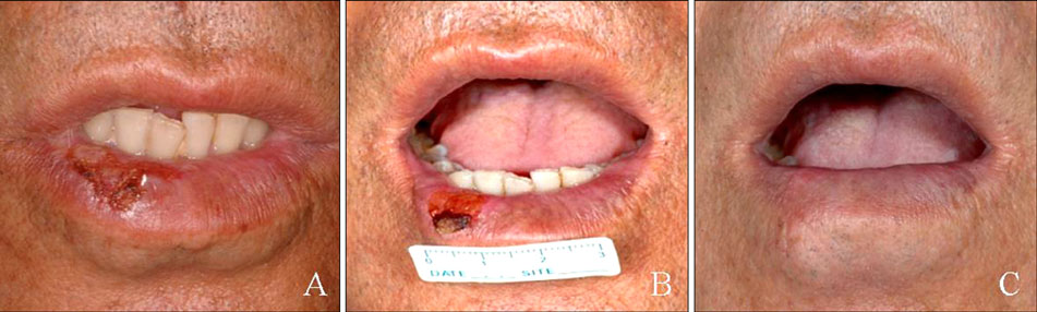
Fig. 1 Clinical presentation of our patient. (A) Initial visit, (B) 30 months later, (C) after excision.

Fig. 2 Histology of our patient. (A) Chronic inflammatory cell infiltration in the dermis without epidermal change at the initial visit. (B) Atypical, pleomorphic keratinocytes are confined to the epidermis 30 months later. (C) Extension of atypical keratinocytes in the tumor nest beyond the basement membrane after excision. (D) Suggestive squamous cell carcinoma in the lymph node (A, B, C, D: H&E, ×40, ×400).

Fig. 3 (A) A whole body computed tomography showing increased signal intensity in the right submandibular area. (B) The positron emission tomography-computed tomography image shows increased fluorine-18-fluorodeoxyglucose (18F-FDG) uptake in the same area, suggestive of lymph node metastasis (arrow).
Reference
-
1. Ducan KO, Geisse JK, Leffell DJ. Wolff K, Goldsmith LA, Katz SI, Gilchrest BA, Paller AS, Leffell DJ, editors. Epithelial precancerous lesions. Fitzpatrick's dermatology in general medicine. 2008. 7th ed. New York: Mc Graw Hill;1007–1027.2. Markopoulos A, Albanidou-Farmaki E, Kayavis I. Actinic cheilitis: clinical and pathologic characteristics in 65 cases. Oral Dis. 2004. 10:212–216.
Article3. Cavalcante AS, Anbinder AL, Carvalho YR. Actinic cheilitis: clinical and histological features. J Oral Maxillofac Surg. 2008. 66:498–503.
Article4. Marks R, Rennie G, Selwood TS. Malignant transformation of solar keratoses to squamous cell carcinoma. Lancet. 1988. 1:795–797.
Article5. Abreu MA, Silva OM, Neto Pimentel DR, Hirata CH, Weckx LL, Alchorne MM, et al. Actinic cheilitis adjacent to squamous carcinoma of the lips as an indicator of prognosis. Braz J Otorhinolaryngol. 2006. 72:767–771.
Article6. Glogau RG. The risk of progression to invasive disease. J Am Acad Dermatol. 2000. 42:23–24.
Article7. Picascia DD, Robinson JK. Actinic cheilitis: a review of the etiology, differential diagnosis, and treatment. J Am Acad Dermatol. 1987. 17:255–264.
Article8. Moy RL. Clinical presentation of actinic keratoses and squamous cell carcinoma. J Am Acad Dermatol. 2000. 42:8–10.
Article9. Robinson JK. Actinic cheilitis. A prospective study comparing four treatment methods. Arch Otolaryngol Head Neck Surg. 1989. 115:848–852.
Article
- Full Text Links
-
- Actions
-
Cited
- CITED
-
- Close
- Share
-
- Similar articles
-
- A Case of squamous Cell Carcinoma Arising from an Actinic Cheilitis and Its Surgical Treatment
- A Case of Actinic Cheilitis Treated by Topical Photodynamic Therapywith Methyl Aminolevulinate
- Pigmented Squamous Cell Carcinoma Arising from Pigmented Actinic Keratosis
- Early Squamous Cell Carcinoma Arising from Disseminated Superficial Actinic Porokeratosis
- A Case of Metastatic Cutaneous Squamous Cell Carcinoma Arising in Chronic Osteomyelitic Focus
