Ann Dermatol.
2014 Apr;26(2):283-285. 10.5021/ad.2014.26.2.283.
Anetoderma Developing in Generalized Granuloma Annulare in an Infant
- Affiliations
-
- 1Department of Dermatology, Hanyang University College of Medicine, Seoul, Korea. tuentuen@hanyang.ac.kr
- KMID: 2171678
- DOI: http://doi.org/10.5021/ad.2014.26.2.283
Abstract
- No abstract available.
MeSH Terms
Figure
-
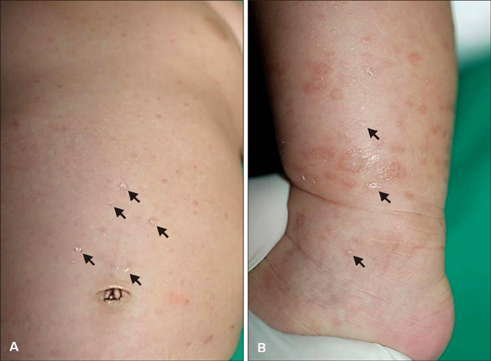
Fig. 1 Multiple atrophic and brownish scaly macules (black arrows) can be seen on the abdomen (A) and left leg (B).
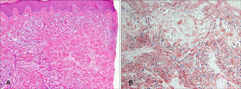
Fig. 2 (A) Skin biopsy showing pale-staining, well-circumscribed, haphazardly arranged degenerated collagen bundles surrounded by inflammatory cells in the dermis (H&E, ×100). (B) Loss of elastic fibers in the superficial and mid dermis (elastic stain, ×200).
Reference
-
1. Yu HJ, Shin H, Kang MS, Kim JS. A case of primary anetoderma in an infant. Br J Dermatol. 2007; 157:1267–1269.
Article2. Güneş P, Göktay F, Mansur AT, Köker F, Erfan G. Collagen-elastic tissue changes and vascular involvement in granuloma annulare: a review of 35 cases. J Cutan Pathol. 2009; 36:838–844.
Article3. Ozkan S, Fetil E, Izler F, Pabucçuoğlu U, Yalçin N, Güneş AT. Anetoderma secondary to generalized granuloma annulare. J Am Acad Dermatol. 2000; 42:335–338.
Article4. Sanyal S, Hejmadi R, Taibjee SM. Granuloma annulare resolving with features of mid-dermal elastolysis. Clin Exp Dermatol. 2009; 34:e1017–e1018.
Article5. Werth VP, Ivanov IE, Nussenzweig V. Decay-accelerating factor in human skin is associated with elastic fibers. J Invest Dermatol. 1988; 91:511–516.
Article
- Full Text Links
-
- Actions
-
Cited
- CITED
-
- Close
- Share
-
- Similar articles
-
- Anetoderma due to Generalized Perforating Granuloma Annulare
- Two Cases of Generalized Granuloma Annulare in Early Childhood
- A Case of Generalized Granuloma Annulare in Infant
- Generalized Actinic Granuloma annulare with Impaired Glucose Tolerance Test
- Three Cases of Generalized Granuloma Annulare in Infancy Following Bacillus Calmette-Guerin Vaccination
