Chromatin organization and transcriptional activation of Hox genes
- Affiliations
-
- 1Department of Anatomy, Embryology Laboratory, Yonsei University College of Medicine, Seoul, Korea. mhkim1@yuhs.ac
- 2Brain Korea 21 Project for Biomedical Science, Yonsei University College of Medicine, Seoul, Korea.
- KMID: 2168900
- DOI: http://doi.org/10.5115/acb.2010.43.1.78
Abstract
- Spatially and temporally programmed expression of the Hox genes along the antero-posterior (A-P) axis is essential for correct pattern formation during embryonic development. An accumulating body of evidence indicates the pivotal role of spatial chromatin organization for the coordination of gene regulation. Recently, chromosome conformation capture (3C) technique has been developed and opened a new way to study chromosomal interactions in the nucleus. In this study, we describe 3C method we applied in F9 embryonic teratocarcinoma cells and demonstrate that the chromosomal interactions at Hox loci are successfully detected. Interestingly, at Hoxc loci, the abundance of intrachromosomal interactions with neighboring fragments was drastically decreased when the genes are expressed. These results indicate the possibility of the dynamic pattern of chromosomal interaction in association with the transcriptional regulation of Hox genes.
Keyword
MeSH Terms
Figure
-
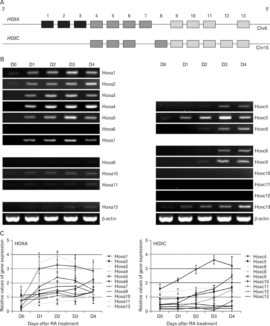
Fig. 1 Expression pattern of Hoxa and Hoxc cluster genes following RA treatment. (A) Hoxa and Hoxc gene organization in each cluster. (B) F9 cells were cultured with RA for 4 days. Total RNAs were isolated each day from Day 1 to Day 4 (D0: control cells with no RA treatment), and then RT-PCR was performed with each Hox primers. β-actin was used as a positive control for amplifiable cDNA. Data are representative of one of three replicate experiments. (C) The relative expression level of each Hox gene was determined by using Multi Gauge software. The amount of each RT-PCR product was normalized with that of β-actin to obtain relative intensities. Each point represents the average of three separate experiments. Error bars represent SEM.

Fig. 2 PCR analysis of restriction digestion and ligation efficiency used for the evaluation of 3C template (A) To determine digestion efficiency, primer pairs D1/D2 and D3/D4 were used. When the HindIII restriction digestion was completed, D1/D2 primer pair gives a faint PCR band whereas D3/D4 primers generate a band whether DNA templates are undigested (-) and digested (+) with HindIII. (B) To determine the efficiency of self ligation, the primer L1/L2 was used. DNA was prepared before (-) and after (+) the ligation step. The 3C templates from Day 3 of RA treatment were also tested with same ways and obtained similar results (Data not shown).
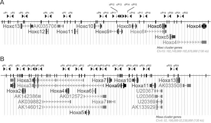
Fig. 3 Schematic presentation of the Hoxc (A) and Hoxa (B) loci showing the primer as well as HindIII regcognition sites. The UCSC Genes track includes both protein-coding and putative non-coding transcripts. The black bar depicts the HindIII recognition sites. The position of each primer we used for 3C analysis is indicated as arrowhead along with a serial number (cP1-cP22 and aP1-aP24 in Hoxc and Hoxa clusters, respectively). Each Hoxc and Hoxa region we analyzed covers 130 kb in mouse chromosome 15 and 6, respectively.
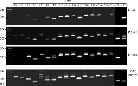
Fig. 4 Identification of intrachromosomal interaction of RA-untreated (D0) samples at Hoxc loci. 3C-PCR was performed with the primers indicated. The primer cP12 was used as an anchor. 3C DNA templates of 250 ng were used per each PCR. This experiment was performed three times (D0-#1, D0-#2, D0-#3) for validation of this method.
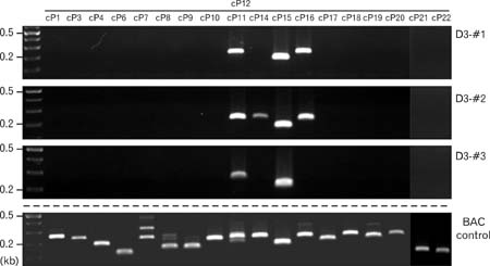
Fig. 5 Identification of intrachromosomal interaction of RA-treated (D3) samples at Hoxc loci. 3C-PCR was performed with the primers indicated. The primer cP12 was used as an anchor. 3C DNA templates of 250 ng were used per each PCR. This experiment was performed three times (D3-#1, D3-#2, D3-#3) for validation. Compare to those of Fig. 4, many PCR bands were disappeared indicating the disappearance of intrachromosomal interaction.
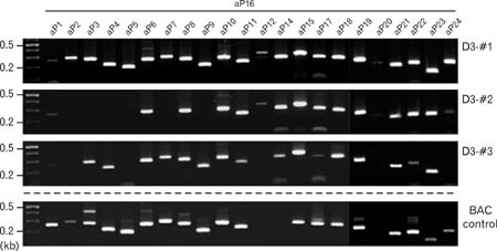
Fig. 6 Identification of intrachromosomal interaction of RA-treated (D3) samples at Hoxa loci. 3C-PCR was performed with the primers indicated. The primer aP16 was used as an anchor. 3C DNA templates of 250 ng were used per each PCR. This experiment was performed three times (D3-#1, D3-#2, D3-#3) for validation.
Reference
-
1. Boncinelli E, Simeone A, Acampora D, Mavilio F. HOX gene activation by retinoic acid. Trends Genet. 1991. 7:329–334.2. Boney-Montoya J, Ziegler YS, Curtis CD, Montoya JA, Nardulli AM. Long-Range transcriptional control of progesterone receptor gene expression. Mol Endocrinol. 2010. 24:346–358.3. Breier G, Bućan M, Francke U, Colberg-Poley AM, Gruss P. Sequential expression of murine homeo box genes during F9 EC cell differentiation. EMBO J. 1986. 5:2209–2215.4. Carter D, Chakalova L, Osborne CS, Dai YF, Fraser P. Long-range chromatin regulatory interactions in vivo. Nat Genet. 2002. 32:623–626.5. Chambeyron S, Bickmore WA. Chromatin decondensation and nuclear reorganization of the HoxB locus upon induction of transcription. Genes Dev. 2004. 18:1119–1130.6. Chavanas S, Adoue V, Mechin MC, et al. Long-range enhancer associated with chromatin looping allows AP-1 regulation of the peptidylarginine deiminase 3 gene in differentiated keratinocyte. PLoS ONE. 2008. 3:e3408.7. Dekker J. Ggene regulation in the third dimension. Science. 2008. 319:1793–1794.8. Dekker J, Rippe K, Dekker M, Kleckner N. Capturing chromosome conformation. Science. 2002. 295:1306–1311.9. Fraser J, Rousseau M, Shenker S, et al. Chromatin conformation signatures of cellular differentiation. Genome Biology. 2009. 10:R37.10. Gaunt SJ, Strachan L. Temporal colinearity in expression of anterior hox genes in developing chick embryos. Dev Dyn. 1996. 207:270–280.11. Izpisúa-Belmonte JC, Falkenstein H, Dollé P, Renucci A, Duboule D. Murine genes related to the Drosophila AbdB homeotic genes are sequentially expressed during development of the posterior part of the body. EMBO J. 1991. 10:2279–2289.12. Kleinjan DA, Lettice LA, Veronica van H, Robert EH. Chapter 13 long range gene control and genetic disease. Advances in genetics, academic press. 2008. Volume 61:339–388.13. Lomvardas S, Barnea G, Pisapia DJ, Mendelsohn M, Kirkland J, Axel R. Interchromosomal interactions and olfactory receptor choice. Cell. 2006. 126:403–413.14. Murphy SP, Garbern J, Odenwald WF, Lazzarini RA, Linney E. Differential expression of the homeobox gene Hox-1.3 in F9 embryonal carcinoma cells. Proc Natl Acad Sci USA. 1988. 85:5587–5591.15. Murrell A, Heeson S, Reik W. Interaction between differentially methylated regions partitions the imprinted genes Igf2 and H19 into parent-specific chromatin loops. Nat Genet. 2004. 36:889–893.16. Spilianakis CG, Flavell RA. Long-range intrachromosomal interactions in the T helper type 2 cytokine locus. Nat Immunol. 2004. 5:1017–1027.17. Stornaiuolo A, Acampora D, Pannese M, et al. Human HOX genes are differentially activated by retinoic acid in embryonal carcinoma cells according to their position within the four loci. Cell Differ Dev. 1990. 31:119–127.18. Tolhuis B, Palstra R-J, Splinter E, Grosveld F, de Laat W. Looping and interaction between hypersensitive sites in the active beta-globin locus. Mol Cell. 2002. 10:1453–1465.19. Wang KC, Helms JA, Chang HY. Regeneration, repair and remembering identity: the three Rs of Hox gene expression. Trends Cell Biol. 2009. 19:268–275.20. Würtele H, Chartrand P. Genome-wide scanning of HoxB1-associated loci in mouse ES cells using an open-ended chromosome conformation capture methodology. Chromosome Res. 2006. 14:477–495.21. Yu SJ, Lee JY, Kim SH, Deocaris CC, Kim MH. Synthetic maternal stress hormone can modulate the expression of Hox genes. J Exp Biomed Sci. 2009. 15:249–255.
- Full Text Links
-
- Actions
-
Cited
- CITED
-
- Close
- Share
-
- Similar articles
-
- Influence of Polycomb Proteins and Epigenetic Transcriptional Modifiers on the Development and Activation of T Lymphocytes
- The Function of the Vitamin D Receptor and a Possible Role of Enhancer RNA in Epigenomic Regulation of Target Genes: Implications for Bone Metabolism
- Epigenetics and Psychiatric Disorders
- Effects of estrogen receptor and estrogen on the chromatin structure in estrogen receptor stable transfectants
- Epigenetic regulation and chromatin remodeling in learning and memory
