Successful Chemotherapy Following Autologous Stem Cell Transplantation in Multiple Myeloma and Multi-organ Dysfunction with Infiltration of Eosinophils: A Case Report
- Affiliations
-
- 1Department of Internal Medicine, Kosin University College of Medicine, Busan, Korea. towersue@hotmail.com
- 2Department of Pathology, Kosin University College of Medicine, Busan, Korea.
- 3Department of Radiology, Kosin University College of Medicine, Busan, Korea.
- 4Department of Laboratory Medicine, Kosin University College of Medicine, Busan, Korea.
Abstract
- Eosinophils are derived from hematopoietic stem cells. Peripheral blood eosinophilia is defined as an absolute eosinophil count of > or =0.5x10(9)/L. Eosinophilia is classified into primary or clonal eosinophilia, secondary eosinophilia, and idiopathic categories including idiopathic hypereosinophilic syndrome. Both hematopoietic and solid neoplasms may be associated with peripheral blood eosinophilia, but multiple myeloma is rarely associated with eosinophilia. We now report the case of a 31-year-old man with multiple myeloma associated with marked eosinophilia who developed multiple organ dysfunction with infiltration of eosinophils. He recovered after treatment with chemotherapy followed by autologous stem cell transplantation.
MeSH Terms
Figure
-

Fig. 1 (A) A bone marrow biopsy showed hypercellular marrow (>90%) with numerous eosinophils and plasma cells (H&E staining,×1,000). (B) Bone marrow aspiration revealed hypercellular marrow with many eosinophils and plasma cells (Wright staining,×1,000).
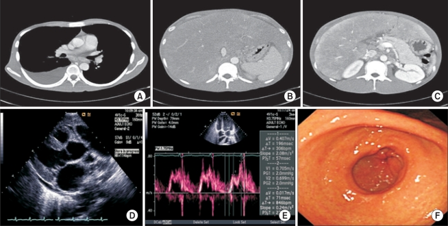
Fig. 2 (A) A right pleural effusion was noted, with focal consolidation or subsegmental atelectasis of the right middle lobe. (B, C) Hepatosplenomegaly of both hepatic lobes was present without a definite focal lesion. The perihepatic space contained fluid. The lymph nodes were enlarged in the peripancreatic, mesenteric and aortocaval areas. (D) Minimal pericardial effusion, septal hypertrophy and mild global hypokinesia of the left ventricle were noted on echocardiography. (E) There was a relaxation abnormality in the mitral valve inflow pattern. (F) Diffuse-fashioned erythema of the mucosa was noted in the antrum of the stomach.

Fig. 3 (A) The liver parenchyme showed infiltrations of numerous eosinophils and plasma cells on the hemorrhage focus (H&E staining,×200). (B) The liver parenchyme and portal areas showed infiltration of eosinophils and plasma cells on the hemorrhage foci (H&E staining,×400). (C, D) Plasma cells and eosinophils were observed in the pleural fluid and the ascitic fluid (D) (H&E staining, C,×1,000; D,×1,000).
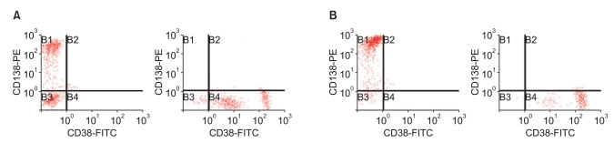
Fig. 4 Flow cytometric detection of plasma cells. Identification of plasma cells in pleural and peritoneal fluids of the patient using the plasma cell markers CD38 and CD138. These cells are high-intensity CD38-positive and CD138-positive. (A) Plasma cell markers were positive (CD138, 60.12%; CD38, 96.2%) in the peritoneal fluid. (B) Plasma cell markers were positive (CD138, 96.18%; CD38, 99.8%) in the pleural fluid.
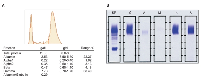
Fig. 5 (A) A monoclonal peak is observed in the gammaglobulin fraction (7.73 g/dL) in serum protein electrophoresis. (B) IgG (G), kappa (κ), and lambda (λ) lanes were identified in serum immunofixation electrophoresis. SP, standard protein; A, IgA; M, IgM.
Reference
-
1. Denburg JA, Telizyn S, Messner H, Lim B, Jamal N, Ackerman SJ, et al. Heterogeneity of human peripheral blood eosinophil-type colonies: evidence for a common basophileosinophil progenitor. Blood. 1985; 66:312–318. PMID: 2410064.
Article2. Valent P. Pathogenesis, classification, and therapy of eosinophilia and eosinophil disorders. Blood Rev. 2009; 23:157–165. PMID: 19246139.
Article3. Tefferi A, Patnaik MM, Pardanani A. Eosinophilia: secondary, clonal and idiopathic. Br J Haematol. 2006; 133:468–492. PMID: 16681635.
Article4. Tefferi A. Blood eosinophilia: a new paradigm in disease classification, diagnosis, and treatment. Mayo Clin Proc. 2005; 80:75–83. PMID: 15667033.
Article5. Samoszuk M. Eosinophils and human cancer. Histol Histopathol. 1997; 12:807–812. PMID: 9225164.6. Franchi F, De Rosa F, Seminara P, Calvieri S, Carfagna GF, Bosman C. Hypereosinophilic syndrome and plasmocytoma. Report of a case and review of the literature. Acta Haematol. 1984; 72:14–20. PMID: 6433626.7. Glantz L, Rintels P, Samoszuk M, Medeiros LJ. Plasma cell myeloma associated with eosinophilia. Am J Clin Pathol. 1995; 103:583–587. PMID: 7741103.
Article8. Kim DW, Shin MG, Yun HK, Kim SH, Shin JH, Suh SP, et al. Incidence and causes of hypereosinophilia (corrected) in the patients of a university hospital. Korean J Lab Med. 2009; 29:185–193. PMID: 19571614.9. Roufosse FE, Goldman M, Cogan E. Hypereosinophilic syndromes. Orphanet J Rare Dis. 2007; 2:37. PMID: 17848188.
Article10. Desenne JJ, Acquatella G, Stern R, Muller A, Sanchez M, Somoza R. Blood eosinophilia in Hodgkin's disease: a follow-up of 25 cases in Venezuela. Cancer. 1992; 69:1248–1253. PMID: 1739923.
Article11. Navarro-Román L, Medeiros LJ, Kingma DW, Zárate-Osorno A, Nguyen V, Samoszuk M, et al. Malignant lymphomas of B-cell lineage with marked tissue eosinophilia: a report of five cases. Am J Surg Pathol. 1994; 18:347–356. PMID: 8141429.12. Lakatos PL, Fekete S, Horanyi M, Fischer S, Abonyi ME. Development of multiple myeloma in a patient with chronic hepatitis C: a case report and review of the literature. World J Gastroenterol. 2006; 12:2297–2300. PMID: 16610042.
Article13. Stefanini M, Claustro JC, Motos RA, Bendigo LL. Blood and bone marrow eosinophilia in malignant tumors: role and nature of blood and tissue eosinophil colony-stimulating factor(s) in two patients. Cancer. 1991; 68:543–548. PMID: 2065275.
Article
- Full Text Links
-
- Actions
-
Cited
- CITED
-
- Close
- Share
-
- Similar articles
-
- HBV reactivation in a HBsAg-negative patient with multiple myeloma treated with prednisolone maintenance therapy after autologous HSCT
- A Case of Cerebral Toxoplasmosis Following Tandem Autologous Stem Cell Transplantation in a Multiple Myeloma Patient
- A case of subcutaneous plasmacytomas of vessel puncture site in multiple myeloma after autologous stem cell transplantation
- Emergence of chronic myeloid leukemia following autologous stem cell transplantation in a young woman with multiple myeloma
- Complete Remission in a Patient with Multiple Myeloma after High Dose Melphalan and CD34+ Selected Autologous Transplantation
