Cancer Res Treat.
2010 Dec;42(4):239-243.
A Case of Blastic Plasmacytoid Dendritic Cell Neoplasm Initially Mimicking Cutaneous Lupus Erythematosus
- Affiliations
-
- 1Department of Internal Medicine, Inha University Hospital, Incheon, Korea. cskimmd@inha.ac.kr
- 2Department of Dermatology, Inha University Hospital, Incheon, Korea.
- 3Department of Pathology, Inha University Hospital, Incheon, Korea.
- 4Department of Laboratory Medicine, Inha University Hospital, Incheon, Korea.
Abstract
- Blastic plasmacytoid dendritic cell neoplasm (BPDCN) is a rare disease. The prognosis is poor in most cases with rapid progression despite administering chemotherapy. A 67-year-old man complained of skin rashes on his back and this spread to the trunk, face, arms and thighs, and he was initially diagnosed with cutaneous lupus erythematosus according to the skin biopsy. The skin rashes then became aggravated on a trial of low dose methylprednisolone for 3 months. Repeated skin biopsy revealed a diffuse infiltration of lymphoid cells with medium sized nuclei, positive for CD4 and CD56, negative for Epstein-Barr virus (EBV), indicating a diagnosis of BPDCN. Further workups confirmed stage IVA BPDCN involving the skin, multiple lymph nodes, the peripheral blood and the bone marrow. He was treated with six cycles of combination chemotherapy consisting of ifosphamide, methotrexate, etoposide, prednisolone and L-asparaginase, and he achieved a partial response. Herein we report on a rare case of BPDCN that was initially misinterpreted as cutaneous lupus erythematosus.
MeSH Terms
Figure
-

Fig. 1 Multiple nodules and plaques on the (A) cheek and (B) trunk.
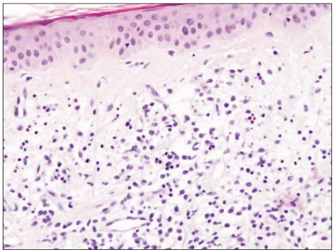
Fig. 2 Findings of the initial skin biopsy. It shows scattered karyorrhectic debris and eosinophilic swelling of the dermal collagen fibers with a lymphocytic infiltration (×200).
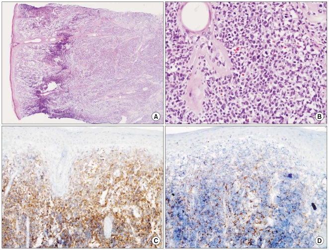
Fig. 3 Findings of the second skin biopsy. (A) Shows an extensive diffuse infiltration of lymphoid cells (H&E, ×40). (B) Shows medium to large size lymphoid cells with nuclear atypia (H&E, ×200). (C) Shows the positive staining for CD4 and (D) shows the positive stain for CD56 on immunohistochemistry (×100).
Reference
-
1. Adachi M, Maeda K, Takekawa M, Hinoda Y, Imai K, Sugiyama S, et al. High expression of CD56 (N-CAM) in a patient with cutaneous CD4-positive lymphoma. Am J Hematol. 1994; 47:278–282. PMID: 7526680.
Article2. Bueno C, Almeida J, Lucio P, Marco J, Garcia R, de Pablos JM, et al. Incidence and characteristics of CD4(+)/HLA DRhi dendritic cell malignancies. Haematologica. 2004; 89:58–69. PMID: 14754607.3. Oshimi K, Kawa K, Nakamura S, Suzuki R, Suzumiya J, Yamaguchi M, et al. NK-cell neoplasms in Japan. Hematology. 2005; 10:237–245. PMID: 16019472.
Article4. Kim Y, Kang MS, Kim CW, Sung R, Ko YH. CD4+CD56+ lineage negative hematopoietic neoplasm: so called blastic NK cell lymphoma. J Korean Med Sci. 2005; 20:319–324. PMID: 15832009.5. Lee M, Ko Y, Kim K, Kim M, Kim M, Cho K. Clinicopathologic and genetic study of cutaneous blastic NK lymphoma in Korea. Korean J Dermatol. 2007; 45(suppl 2):145.6. Petrella T, Dalac S, Maynadié M, Mugneret F, Thomine E, Courville P, et al. Groupe Français d'Etude des Lymphomes Cutanés (GFELC). CD4+CD56+ cutaneous neoplasms: a distinct hematological entity? Am J Surg Pathol. 1999; 23:137–146. PMID: 9989839.7. Willemze R, Jaffe ES, Burg G, Cerroni L, Berti E, Swerdlow SH, et al. WHO-EORTC classification for cutaneous lymphomas. Blood. 2005; 105:3768–3785. PMID: 15692063.
Article8. Chaperot L, Bendriss N, Manches O, Gressin R, Maynadie M, Trimoreau F, et al. Identification of a leukemic counterpart of the plasmacytoid dendritic cells. Blood. 2001; 97:3210–3217. PMID: 11342451.
Article9. Jaye DL, Geigerman CM, Herling M, Eastburn K, Waller EK, Jones D. Expression of the plasmacytoid dendritic cell marker BDCA-2 supports a spectrum of maturation among CD4+ CD56+ hematodermic neoplasms. Mod Pathol. 2006; 19:1555–1562. PMID: 16998465.
Article10. Cota C, Vale E, Viana I, Requena L, Ferrara G, Anemona L, et al. Cutaneous manifestations of blastic plasmacytoid dendritic cell neoplasm-morphologic and phenotypic variability in a series of 33 patients. Am J Surg Pathol. 2010; 34:75–87. PMID: 19956058.
Article11. Rönnblom L, Pascual V. The innate immune system in SLE: type I interferons and dendritic cells. Lupus. 2008; 17:394–399. PMID: 18490415.
Article12. Jegalian AG, Facchetti F, Jaffe ES. Plasmacytoid dendritic cells: physiologic roles and pathologic states. Adv Anat Pathol. 2009; 16:392–404. PMID: 19851130.13. Vermi W, Lonardi S, Morassi M, Rossini C, Tardanico R, Venturini M, et al. Cutaneous distribution of plasmacytoid dendritic cells in lupus erythematosus. Selective tropism at the site of epithelial apoptotic damage. Immunobiology. 2009; 214:877–886. PMID: 19625100.
Article
- Full Text Links
-
- Actions
-
Cited
- CITED
-
- Close
- Share
-
- Similar articles
-
- A Woman with Blastic Plasmacytoid Dendritic Cell Neoplasm
- Blastic Plasmacytoid Dendritic Cell Neoplasm Mimicking Traumatic Hematoma: A Case Report
- A Case of Blastic Plasmacytoid Dendritic Cell Neoplasm in Child
- A Case of Blastic Plasmacytoid Dendritic Cell Neoplasm with Mutations in DNMT3A, TET2, SRSF2, and ATRX Genes
- Plasmacytoid dendritic cell neoplasms
