Craniofacial Asymmetry in Adults With Neglected Congenital Muscular Torticollis
- Affiliations
-
- 1The Clinic for Torticollis, Department of Physical Medicine and Rehabilitation, Ajou University School of Medicine, Suwon, Korea. syyim@ajou.ac.kr
- KMID: 2165648
- DOI: http://doi.org/10.5535/arm.2015.39.3.440
Abstract
OBJECTIVE
To evaluate the craniofacial asymmetry in adults with neglected congenital muscular torticollis (CMT) by quantitative assessment based on craniofacial three-dimensional computed tomography (3D-CT).
METHODS
Preoperative craniofacial asymmetry was measured by 3D-CT for 31 CMT subjects > or =18 years of age who visited a tertiary medical center and underwent 3D-CT between January 2009 and December 2013. The relationship between the age and the severity of craniofacial asymmetry was analyzed in reference to anteroposterior length asymmetry of the frontal bone and zygomatic arch, vertical and lateral displacements of the facial landmarks, and mandibular axis rotation.
RESULTS
The age at CT was 27.71+/-7.02 years (range, 18-44 years). All intra-class correlation coefficients were higher than 0.7, suggesting good inter-rater reliability (p<0.05) of all the measurements. The frontal and the zygomatic length ratio (i.e., the anteroposterior length asymmetry on the axial plane) was 1.06+/-0.03 and 1.07+/-0.03, respectively, which was increased significantly with age in the linear regression analysis (r2=0.176, p=0.019 and r2=0.188, p=0.015, respectively). The vertical or lateral displacement of the facial landmarks and rotation of the mandibular axis did not significantly correlate with age (p>0.05).
CONCLUSION
Craniofacial asymmetry of neglected CMT became more severe with age in terms of anteroposterior length asymmetry of the ipsilateral frontal bone and zygomatic arch on the axial plane even after growth cessation. This finding may enhance the understanding of therapeutic strategies for craniofacial asymmetry in adults with neglected CMT.
Keyword
MeSH Terms
Figure
-

Fig. 1 Measurement of the anteroposterior length asymmetry on the axial plane. (A) The FLR, b/a, which represents the anteroposterior length asymmetry of the OS on the CMT side in comparison with that on the non-CMT side on the superior cranial view. The z1-axis was defined as the coronal line perpendicular to the sagittal plane, which crosses the intersection of the 2 diagonals from the OS. (B) The ZLR, d/c, which represents the anteroposterior length asymmetry of the ZM on the CMT side in comparison with that on the non-CMT side on the inferior cranial view. The z2-axis was defined as the line connecting the bilateral MA. (C) Linear regression analysis between FLR and the subjects' ages. (D) Linear regression analysis between ZLR and the subjects' ages. The scatter plots of FLR and ZLR show statistically significant linear correlation (*p<0.05) with age. OS, orbitale superius; CMT, congenital muscular torticollis; ZM, zygomaxillare; MA, mastoid processes.
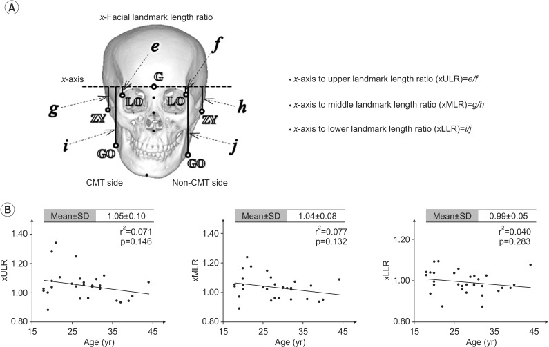
Fig. 2 Measurement of the vertical length asymmetry on the coronal plane. (A) A mid-sagittal line was constructed by joining the landmarks, namely the glabella 'G', nasion 'N', and top of the nasal spine 'TNS', to form the y1-axis and perpendicular line with origins at G, defined as the x-axis on the frontal view. The xULR, e/f, represents the vertical length asymmetry of the LO from the x-axis on the CMT side in comparison with that on the non-CMT side. The xMLR, g/h, represents the vertical length asymmetry of the ZY. The xLLR, i/j, represents the vertical length asymmetry of the GO on the CMT side in comparison with that on the non-CMT side. (B) Linear regression analyses between xULR, xMLR, xLLR, and the subjects' ages. LO, latero-orbitale; CMT, congenital muscular torticollis; ZY, zygonion; GO, gonion.
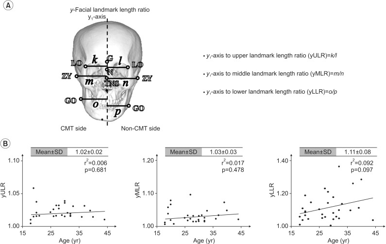
Fig. 3 Measurement of the lateral length asymmetry on the coronal plane. (A) The distances from the y1-axis to the paired landmarks mentioned in Fig. 2A were measured in frontal view. The yULR, k/l, which represents the lateral length asymmetry of the LO from the y1-axis on the CMT side in comparison with that on the non-CMT side. The yMLR, m/n, represents the lateral length asymmetry of ZY. The yLLR, o/p, represents the lateral length asymmetry of GO on the CMT side in comparison with that on the non-CMT side. (B) Linear regression analyses between yULR, yMLR, yLLR, and the subjects' ages. LO, latero-orbitale; CMT, congenital muscular torticollis; ZY, zygonion; GO, gonion.
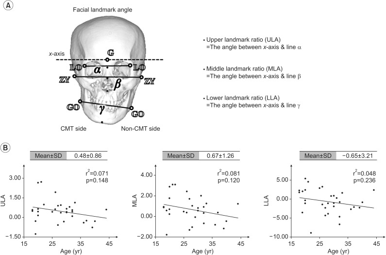
Fig. 4 Measurement of the vertical displacement on the coronal plane. (A) The ULA, which represents the vertical displacement of the LO on the CMT side in comparison with that on the non-CMT side, is the angle between the x-axis and line α. The MLA, which represents the vertical displacement of the ZY on the CMT side in comparison with that on the non-CMT side, is the angle between the x-axis and line β. The ULA, which represents the vertical displacement of the GO on the CMT side in comparison with that on the non-CMT side, is the angle between the x-axis and line γ. (B) Linear regression analyses between ULA, MLA, LLA, and the subjects' ages. LO, latero-orbitale; CMT, congenital muscular torticollis; ZY, zygonion; GO, gonion.
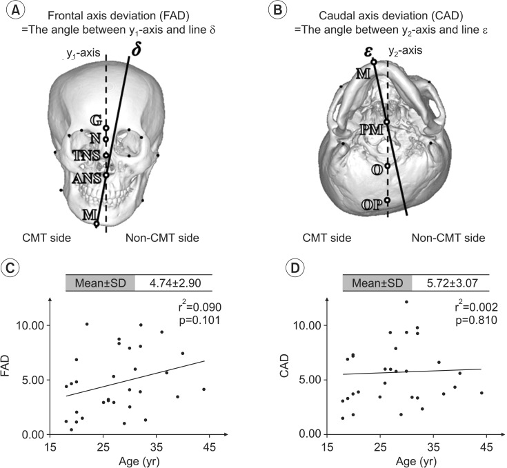
Fig. 5 Measurement of the mandibular axis rotation. (A) The FAD is the angle formed by the intersection of the y1-axis and line δ, which represents the frontal mandibular axis, defined by a line connecting the nasal spine (ANS) and M on the frontal view. (B) The CAD is the angle between the y2-axis and line ε, which represents the caudal mandibular axis. The y2-axis was defined by joining the landmarks OP and O. The caudal mandibular axis was constructed by joining the landmarks M, PM in the inferior cranial view. (C) Linear regression analysis between FAD and the subjects' ages. (D) Linear regression analysis between CAD and the subjects' ages. ANS, anterior nasal spine; TNS, top of the nasal spine; M, mentum; OP, occipital protuberance; O, opisthion; PM, palatine midpoint; G, glabella; N, nasion.
Cited by 2 articles
-
Effectiveness of Surgical Release in Patients With Neglected Congenital Muscular Torticollis According to Age at the Time of Surgery
Kyung-Jay Min, Ah-Reum Ahn, Eun-Ji Park, Shin-Young Yim
Ann Rehabil Med. 2016;40(1):34-42. doi: 10.5535/arm.2016.40.1.34.Occlusal deviations in adolescents with idiopathic and congenital scoliosis
Hao Zhang, Jingbo Ma, Zhicheng Zhang, Yafei Feng, Chuan Cai, Chao Wang
Korean J Orthod. 2022;52(3):165-171. doi: 10.4041/kjod21.259.
Reference
-
1. Cheng JC, Wong MW, Tang SP, Chen TM, Shum SL, Wong EM. Clinical determinants of the outcome of manual stretching in the treatment of congenital muscular torticollis in infants: a prospective study of eight hundred and twenty-one cases. J Bone Joint Surg Am. 2001; 83A:679–687. PMID: 11379737.2. Chen MM, Chang HC, Hsieh CF, Yen MF, Chen TH. Predictive model for congenital muscular torticollis: analysis of 1021 infants with sonography. Arch Phys Med Rehabil. 2005; 86:2199–2203. PMID: 16271571.
Article3. Cheng JC, Tang SP, Chen TM, Wong MW, Wong EM. The clinical presentation and outcome of treatment of congenital muscular torticollis in infants: a study of 1,086 cases. J Pediatr Surg. 2000; 35:1091–1096. PMID: 10917303.4. Yim SY, Yoon D, Park MC, Lee IJ, Kim JH, Lee MA, et al. Integrative analysis of congenital muscular torticollis: from gene expression to clinical significance. BMC Med Genomics. 2013; 6(Suppl 2):S10. PMID: 23819832.
Article5. Seo SJ, Yim SY, Lee IJ, Han DH, Kim CS, Lim H, et al. Is craniofacial asymmetry progressive in untreated congenital muscular torticollis? Plast Reconstr Surg. 2013; 132:407–413. PMID: 23584628.
Article6. Lee DY, Song BW, Cho TJ, Choi IH, Chung CY, Yoo WJ. Craniofacial Asymmetry in Congenital Muscular Torticollis Patients: A Study using Cephalometry. J Korean Orthop Assoc. 2007; 42:24–31.7. Yu CC, Wong FH, Lo LJ, Chen YR. Craniofacial deformity in patients with uncorrected congenital muscular torticollis: an assessment from three-dimensional computed tomography imaging. Plast Reconstr Surg. 2004; 113:24–33. PMID: 14707619.
Article8. Lim KS, Shim JS, Lee YS. Is sternocleidomastoid muscle release effective in adults with neglected congenital muscular torticollis? Clin Orthop Relat Res. 2014; 472:1271–1278. PMID: 24258687.
Article9. Hwang JH, Lee HB, Kim JH, Park MC, Kwack KS, Han JD, Yim SY. Magnetic resonance imaging as a determinant for surgical release of congenital muscular torticollis: correlation with the histopathologic findings. Ann Rehabil Med. 2012; 36:320–327. PMID: 22837966.
Article10. Shim JS, Jang HP. Operative treatment of congenital torticollis. J Bone Joint Surg Br. 2008; 90:934–939. PMID: 18591606.
Article11. Chate RA. Facial scoliosis due to sternocleidomastoid torticollis: a cephalometric analysis. Int J Oral Maxillofac Surg. 2004; 33:338–343. PMID: 15145034.
Article12. Lee JK, Moon HJ, Park MS, Yoo WJ, Choi IH, Cho TJ. Change of craniofacial deformity after sternocleidomastoid muscle release in pediatric patients with congenital muscular torticollis. J Bone Joint Surg Am. 2012; 94:e93. PMID: 22760394.
Article13. Wilbrand JF, Wilbrand M, Malik CY, Howaldt HP, Streckbein P, Schaaf H, et al. Complications in helmet therapy. J Craniomaxillofac Surg. 2012; 40:341–346. PMID: 21741852.
Article14. Loveday BP, de Chalain TB. Active counterpositioning or orthotic device to treat positional plagiocephaly? J Craniofac Surg. 2001; 12:308–313. PMID: 11482615.
Article15. Khoury J. CORR Insights: Is sternocleidomastoid muscle release effective in adults with neglected congenital muscular torticollis? Clin Orthop Relat Res. 2014; 472:1279–1280. PMID: 24307064.
Article16. Tse P, Cheng J, Chow Y, Leung PC. Surgery for neglected congenital torticollis. Acta Orthop Scand. 1987; 58:270–272. PMID: 3630660.
Article17. Oh I, Nowacek CJ. Surgical release of congenital torticollis in adults. Clin Orthop Relat Res. 1978; (131):141–145. PMID: 657610.
Article
- Full Text Links
-
- Actions
-
Cited
- CITED
-
- Close
- Share
-
- Similar articles
-
- Craniofacial Asymmetry in Congenital Muscular Torticollis Patients: A Study using Cephalometry
- Familial Congenital Muscular Torticollis: A Case Report
- Head Tilt and Facial Asymmetry in Congenital Muscular Torticollis
- Congenital Muscular Torticollis Concurrent With Sagittal Synostosis: A Case Report
- Two Cases of Sternomastoid Tumor
