A Fatal Case of Middle East Respiratory Syndrome Corona Virus Infection in South Korea: Chest Radiography and CT Findings
- Affiliations
-
- 1Department of Radiology, Department of Internal Medicine, Yeouido St. Mary's Hospital, The Catholic University of Korea College of Medicine, Seoul, Korea. horrim@catholic.ac.kr
- 2Division of Infectious Diseases, Department of Internal Medicine, Yeouido St. Mary's Hospital, The Catholic University of Korea College of Medicine, Seoul, Korea.
- KMID: 2164820
- DOI: http://doi.org/10.3348/jksr.2016.74.6.407
Abstract
- The outbreak of Middle East Respiratory Syndrome Corona Virus (MERS-CoV) infection in South Korea originated from Saudi Arabia. This virus shows high infectivity, and causes outbreaks of severe febrile respiratory infections in health care-associated settings. Herein, we reported a fatal case of MERS-CoV infection with a focus on the pulmonary radiologic findings. The initial chest computed tomography and radiographs of our patient showed ground-glass opacity in patchy distribution, followed by rapid progression of consolidation and pleural effusion in serial studies.
MeSH Terms
Figure
-
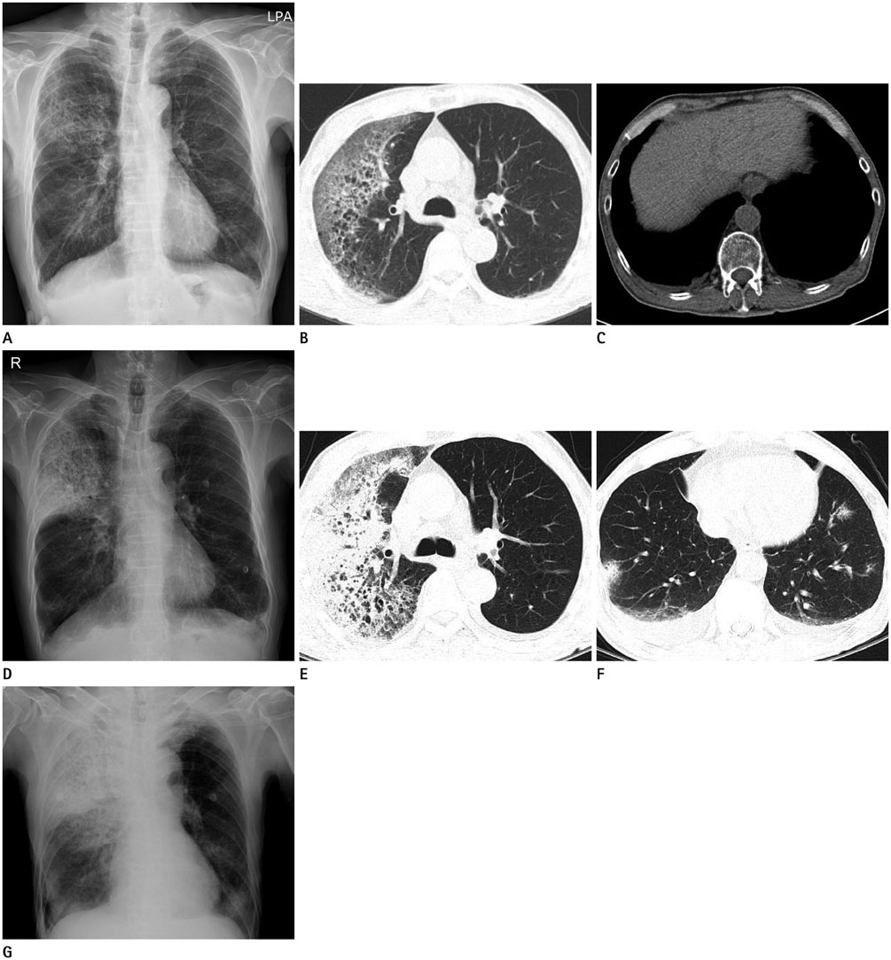
Fig. 1 A 71-year-old man diagnosed with Middle East Respiratory Syndrome Corona Virus. A. The initial chest radiograph shows an ill-defined increased opacity in the right upper lung field. B, C. Chest CT scans obtained on the same day as (A) demonstrate patchy ground-glass opacity and interlobular septal thickenings in the subpleural portion of right upper lobe (B). A small amount of bilateral pleural effusion is also noted (C). D. Follow-up chest radiographs obtained 2 days later reveal progression of consolidation in right upper lung field and newly developed, ill-defined, increased opacity in both lower lung fields. E, F. Follow-up chest CT scans obtained on the same day as (D) show markedly increased extent of consolidation and ground-glass opacity in the right upper lobe (E). Newly developed multifocal nodules in both lower lobes and increased amount of bilateral pleural effusion (F). G. A further follow-up chest radiograph 2 days later, shows aggravated multifocal consolidations in the right upper lung field and both lower lung fields.
Reference
-
1. World Health Organization. Background and summary of novel coronavirus infection - as of 30 November 2012. Accessed Jan 18, 2016. Available at: http://www.who.int/csr/disease/coronavirus_infections/update_20121130/en/.2. Das KM, Lee EY, Enani MA, AlJawder SE, Singh R, Bashir S, et al. CT correlation with outcomes in 15 patients with acute Middle East respiratory syndrome coronavirus. AJR Am J Roentgenol. 2015; 204:736–742.3. World Health Organization. Middle East respiratory syndrome coronavirus (MERS-CoV) - Republic of Korea. Accessed Jan 18, 2016. Available at: http://www.who.int/csr/don/26-june-2015-merskorea/en/.4. Assiri A, Al-Tawfiq JA, Al-Rabeeah AA, Al-Rabiah FA, Al-Hajjar S, Al-Barrak A, et al. Epidemiological, demographic, and clinical characteristics of 47 cases of Middle East respiratory syndrome coronavirus disease from Saudi Arabia: a descriptive study. Lancet Infect Dis. 2013; 13:752–761.5. World Health Organization. Middle East respiratory syndrome coronavirus (MERS-CoV). Accessed Jan 18, 2016. Available at: http://www.who.int/mediacentre/factsheets/mers-cov/en/.6. Majumder MS, Rivers C, Lofgren E, Fisman D. Estimation of MERS-coronavirus reproductive number and case fatality rate for the Spring 2014 Saudi Arabia outbreak: insights from publicly available Data. PLoS Curr. 2014; 12. 18. [Epub]. DOI: 10.1371/currents.outbreaks.98d2f8f3382d84f390736cd5f5fe133c.7. World Health Organization. Summary and risk assessment of current situation in Republic of Korea and China. Accessed Jan 18, 2016. Available at: http://www.who.int/csr/disease/coronavirus_infections/risk-assessment-3june2015/en/.8. Ajlan AM, Ahyad RA, Jamjoom LG, Alharthy A, Madani TA. Middle East respiratory syndrome coronavirus (MERS-CoV) infection: chest CT findings. AJR Am J Roentgenol. 2014; 203:782–787.9. Wong KT, Antonio GE, Hui DS, Lee N, Yuen EH, Wu A, et al. Severe acute respiratory syndrome: radiographic appearances and pattern of progression in 138 patients. Radiology. 2003; 228:401–406.10. Agarwal PP, Cinti S, Kazerooni EA. Chest radiographic and CT findings in novel swine-origin influenza A (H1N1) virus (S-OIV) infection. AJR Am J Roentgenol. 2009; 193:1488–1493.
- Full Text Links
-
- Actions
-
Cited
- CITED
-
- Close
- Share
-
- Similar articles
-
- Middle East Respiratory Syndrome-Coronavirus Infection: A Case Report of Serial Computed Tomographic Findings in a Young Male Patient
- An Unexpected Outbreak of Middle East Respiratory Syndrome Coronavirus Infection in the Republic of Korea, 2015
- The Korean Middle East Respiratory Syndrome Coronavirus Outbreak and Our Responsibility to the Global Scientific Community
- An Atypical Case of Middle East Respiratory Syndrome in a Returning Traveler to Korea from Kuwait, 2018
- 2015 MERS outbreak in Korea: hospital-to-hospital transmission
