Middle East Respiratory Syndrome-Coronavirus Infection: A Case Report of Serial Computed Tomographic Findings in a Young Male Patient
- Affiliations
-
- 1Department of Radiology, Dong-A University Hospital, Busan 49201, Korea. gnlee@dau.ac.kr
- 2Division of Infectious Diseases, Department of Internal Medicine, Dong-A University Hospital, Busan 49201, Korea.
- KMID: 2351177
- DOI: http://doi.org/10.3348/kjr.2016.17.1.166
Abstract
- Radiologic findings of Middle East respiratory syndrome (MERS), a novel coronavirus infection, have been rarely reported. We report a 30-year-old male presented with fever, abdominal pain, and diarrhea, who was diagnosed with MERS. A chest computed tomographic scan revealed rapidly developed multifocal nodular consolidations with ground-glass opacity halo and mixed consolidation, mainly in the dependent and peripheral areas. After treatment, follow-up imaging showed that these abnormalities markedly decreased but fibrotic changes developed.
MeSH Terms
Figure
-
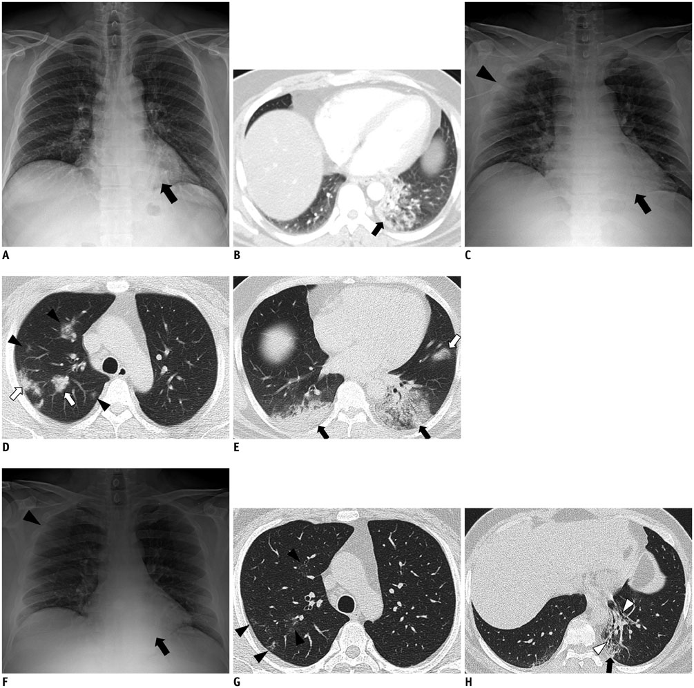
Fig. 1 30-year-old male patient with Middle East respiratory syndrome coronavirus infection. A, B. Initial posteroanterior chest radiograph and abdominal computed tomography (CT) scan were performed in outside hospital on day of admission (6 days after onset of fever). Chest radiograph (A) shows patchy increased opacity (black arrow) in left lower lung zone, retrocardiac area. Axial CT scan (B) shows patchy area of consolidation with air-bronchogram in left lower lobe, which was mainly peripherally located (black arrow). C-E. Follow-up anteroposterior chest radiograph and chest CT scan were taken in outside hospital 4 days after admission (10 days after onset of fever). Chest radiograph (C) shows newly developed patchy area of ill-defined increased opacity in right upper lung zone (black arrowhead) and increased extent of consolidation in left lower lung zone (black arrow). Upper lung CT scan (D) shows multifocal patchy areas of consolidation (white arrows) with ground glass opacity (GGO) halo and nodular GGO lesions (black arrowheads) in both upper lobes, which were mainly slightly peripherally located. Lower lung CT scan (E) shows larger areas of mixed consolidations and GGOs with air-bronchograms (black arrows) in both lower lobes, mainly in dependent area and newly detected focal consolidation in lingular segment of left upper lobe (white arrow). F-H. Last follow-up anteroposterior chest radiograph and chest CT scan were performed on day of discharge (13 days after admission to our hospital; 23 days after onset of fever). Chest radiograph (F) depicts markedly decreased extent of previous consolidations in right upper and left lower lung zones but residual small increased opacity in right upper lung zone (black arrowhead) and left lower lung zone (black arrow). Upper lung CT scan (G) shows markedly reduced extent of previous multifocal patchy areas of consolidation and nodular ground-glass opacity (GGO) lesions in both upper lobes, but also demonstrates residual GGO lesions (black arrowheads). Lower lung CT scan (H) demonstrates markedly decreased extent of previous mixed consolidations and GGOs with air-bronchograms (black arrow), and developed traction bronchiectasis (white arrowheads) with volume loss in left lower lobe, which suggested fibrosis.
Cited by 4 articles
-
Pneumonia Associated with 2019 Novel Coronavirus: Can Computed Tomographic Findings Help Predict the Prognosis of the Disease?
Kyung Soo Lee
Korean J Radiol. 2020;21(3):257-258. doi: 10.3348/kjr.2020.0096.Novel Coronavirus Pneumonia Outbreak in 2019: Computed Tomographic Findings in Two Cases
Xiaoqi Lin, Zhenyu Gong, Zuke Xiao, Jingliang Xiong, Bing Fan, Jiaqi Liu
Korean J Radiol. 2020;21(3):365-368. doi: 10.3348/kjr.2020.0078.Chest Radiographic and CT Findings of the 2019 Novel Coronavirus Disease (COVID-19): Analysis of Nine Patients Treated in Korea
Soon Ho Yoon, Kyung Hee Lee, Jin Yong Kim, Young Kyung Lee, Hongseok Ko, Ki Hwan Kim, Chang Min Park, Yun-Hyeon Kim
Korean J Radiol. 2020;21(4):494-500. doi: 10.3348/kjr.2020.0132.Novel respiratory infectious diseases in Korea
Hyun Jung Kim
Yeungnam Univ J Med. 2020;37(4):286-295. doi: 10.12701/yujm.2020.00633.
Reference
-
1. Zaki AM, van Boheemen S, Bestebroer TM, Osterhaus AD, Fouchier RA. Isolation of a novel coronavirus from a man with pneumonia in Saudi Arabia. N Engl J Med. 2012; 367:1814–1820.2. Ajlan AM, Ahyad RA, Jamjoom LG, Alharthy A, Madani TA. Middle East respiratory syndrome coronavirus (MERS-CoV) infection: chest CT findings. AJR Am J Roentgenol. 2014; 203:782–787.3. Das KM, Lee EY, Enani MA, AlJawder SE, Singh R, Bashir S, et al. CT correlation with outcomes in 15 patients with acute Middle East respiratory syndrome coronavirus. AJR Am J Roentgenol. 2015; 204:736–742.4. Corman VM, Eckerle I, Bleicker T, Zaki A, Landt O, Eschbach-Bludau M, et al. Detection of a novel human coronavirus by real-time reverse-transcription polymerase chain reaction. Euro Surveill. 2012; (17):pii: 20285.5. Alagaili AN, Briese T, Mishra N, Kapoor V, Sameroff SC, Burbelo PD, et al. Middle East respiratory syndrome coronavirus infection in dromedary camels in Saudi Arabia. Mbio. 2014; 5:e00884–e00814. DOI: 10.1128/mbio.00884-14.6. Centers for Disease Control and Prevention. Notice to health care providers: updated guidelines for evaluation of severe respiratory illness associated with Middle East respiratory syndrome coronavirus (MERS-CoV). Accessed June 22, 2015. http://emergency.cdc.gov/HAN/han00348.asp Published June 7, 2013.7. World Health Organization. MERS-CoV in the Republic of Korea at a glance. Accessed June 22, 2015. http://www.wpro.who.int/outbreaks_emergencies/wpro_coronavirus/en/ Published June 22, 2015.8. Assiri A, Al-Tawfiq JA, Al-Rabeeah AA, Al-Rabiah FA, Al-Hajjar S, Al-Barrak A, et al. Epidemiological, demographic, and clinical characteristics of 47 cases of Middle East respiratory syndrome coronavirus disease from Saudi Arabia: a descriptive study. Lancet Infect Dis. 2013; 13:752–761.9. Das KM, Lee EY, Jawder SE, Enani MA, Singh R, Skakni L, et al. Acute Middle East respiratory syndrome coronavirus: temporal lung changes observed on the chest radiographs of 55 patients. AJR Am J Roentgenol. 2015; 205:W267–W274.
- Full Text Links
-
- Actions
-
Cited
- CITED
-
- Close
- Share
-
- Similar articles
-
- An Unexpected Outbreak of Middle East Respiratory Syndrome Coronavirus Infection in the Republic of Korea, 2015
- The Korean Middle East Respiratory Syndrome Coronavirus Outbreak and Our Responsibility to the Global Scientific Community
- A Fatal Case of Middle East Respiratory Syndrome Corona Virus Infection in South Korea: Chest Radiography and CT Findings
- The Same Middle East Respiratory Syndrome-Coronavirus (MERS-CoV) yet Different Outbreak Patterns and Public Health Impacts on the Far East Expert Opinion from the Rapid Response Team of the Republic of Korea
- Middle East Respiratory Syndrome Coronavirus Infection in Children
