Comparison of digital models generated from three-dimensional optical scanner and cone beam computed tomography
- Affiliations
-
- 1Department of Dentistry, School of Dentistry, Seoul National University, Seoul, Republic of Korea.
- 2Department of Oral Microbiology and Immunology, School of Dentistry, Seoul National University, Seoul, Republic of Korea.
- 3Department of Oral and Maxillofacial Radiology, School of Dentistry, Seoul National University, Seoul, Republic of Korea. wjyi@snu.ac.kr
- KMID: 2162380
- DOI: http://doi.org/10.14368/jdras.2016.32.1.60
Abstract
- PURPOSE
The objective of this study was to compare the accuracy of digital models from 3 dimentional (3D) optical scanner and cone beam computed tomography (CBCT).
MATERIALS AND METHODS
We obtained digital models from 11 pairs of stone casts using a 3D optical scanner and a CBCT, and compared the accuracy of the models.
RESULTS
The error range of average positive distance was 0.059 - 0.117 mm and negative distance was 0.066 - 0.146 mm. Statistically (P < 0.05), average positive distance was larger than 70 µm and shorter than 100 µm, and that of negative distance was larger than 100 µm and shorter than 120 µm.
CONCLUSION
We concluded that the accuracy of digital models generated from CBCT is not appropriate to make final prostheses. However, it may be acceptable for provisional restorations and orthodontic diagnoses with respect to the accuracy of the digitalization.
Keyword
MeSH Terms
Figure
-
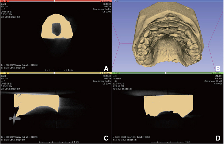
Fig. 1 Construction procedure of digital model from CBCT image. (A) Down view of intermediate model with threshold 600 - 1,000, (B) Rear view of intermediate model with threshold 600 - 1,000, (C) Side view of intermediate model with threshold 600 - 1,000, (D) Constructed digital model.
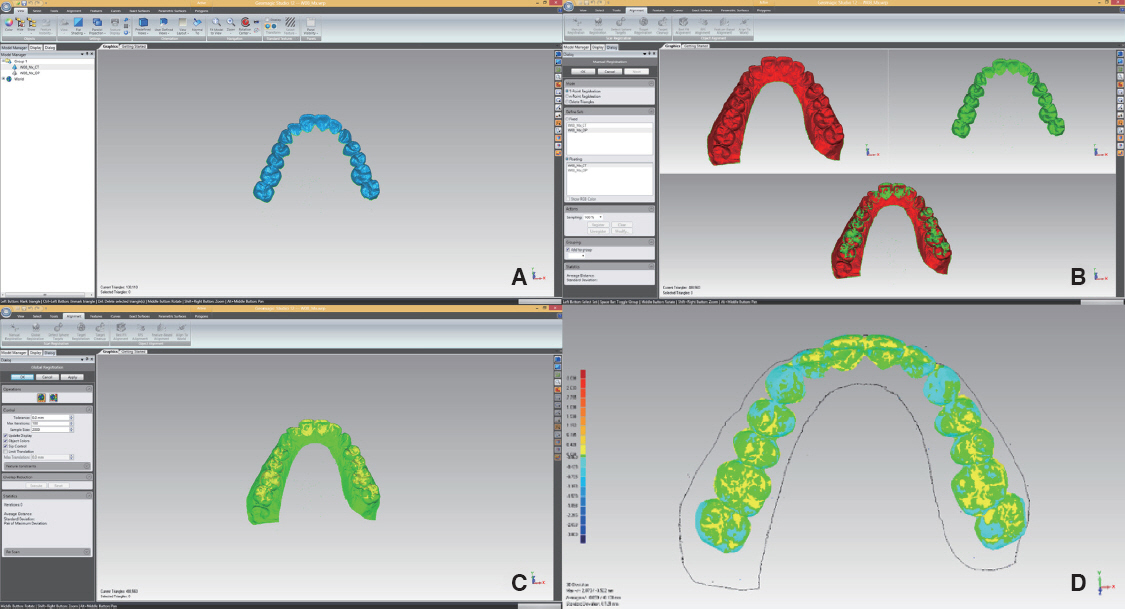
Fig. 2 Deviation analysis procedure. (A) Trimming of CBCT digital model, (B) Alignment of digital models from CBCT and 3D oral scanner by ICP algorithm, (C) Aligned result, (D) Result of deviation analysis.
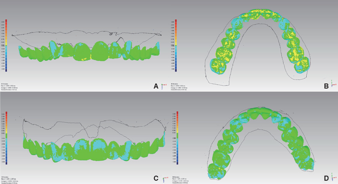
Fig. 3 Deviation analysis (A) Frontal view of result shows least average PE (maxillary cast of model no. 01), (B) Occlusal view of result shows least average PE (maxillary cast of model no. 01), (C) Frontal view of result shows least average NE (maxillary cast of model no. 03), (D) Occlusal view of result shows least average NE (maxillary cast of model no. 03).
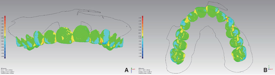
Fig. 4 Deviation analysis which shows PE at pit, fissure, proximal area and NE at cusp (maxillary cast of model no. 10).
Cited by 1 articles
-
Comparison of the accuracy of intraoral scanner by three-dimensional analysis in single and 3-unit bridge abutment model: In vitro study
Mei-Yang Huang, Keunbada Son, Wan-Sun Lee, Kyu-Bok Lee
J Korean Acad Prosthodont. 2019;57(2):102-109. doi: 10.4047/jkap.2019.57.2.102.
Reference
-
References
1. Ender A, Mehl A. Accuracy of complete-arch dental impressions: a new method of measuring trueness and precision. J Prosthet Dent. 2013; 109:121–8. DOI: 10.1016/S0022-3913(13)60028-1.2. Bootvong K, Liu Z, McGrath C, Hagg U, Wong RW, Bendeus M, Yeung S. Virtual model analysis as an alternative approach to plaster model analysis: reliability and validity. Eur J Orthod. 2010; 32:589–95. DOI: 10.1093/ejo/cjp159. PMID: 20164126.3. Birnbaum NS, Aaronson HB. Dental impressions using 3D digital scanners: virtual becomes reality. Compend Contin Educ Dent. 2008; 29:494. 496, 498-505. PMID: 18935788.4. Patzelt SB, Bishti S, Stampf S, Att W. Accuracy of computer-aided design/computer-aided manufacturing-generated dental casts based on intraoral scanner data. J Am Dent Assoc. 2014; 145:1133–40. DOI: 10.14219/jada.2014.87. PMID: 25359645.5. Cuperus AM, Harms MC, Rangel FA, Bronkhorst EM, Schols JG, Breuning KH. Dental models made with an intraoral scanner: a validation study. Am J Orthod Dentofacial Orthop. 2012; 142:308–13. DOI: 10.1016/j.ajodo.2012.03.031. PMID: 22920696.6. Lee CY, Ganz SD, Wong N, Suzuki JB. Use of cone beam computed tomography and a laser intraoral scanner in virtual dental implant surgery: part 1. Implant Dent. 2012; 21:265–71. DOI: 10.1097/ID.0b013e31825e5739. PMID: 22814549.7. Frisardi G, Chessa G, Barone S, Paoli A, Razionale A, Frisardi F. Integration of 3D anatomical data obtained by CT imaging and 3D optical scanning for computer aided implant surgery. BMC Med Imaging. 2011; 11:5. DOI: 10.1186/1471-2342-11-5. PMID: 21338504. PMCID: PMC3047291.8. Swennen GR, Mommaerts MY, Abeloos J, De Clercq C, Lamoral P, Neyt N, Schutyser FA. A cone-beam CT based technique to augment the 3D virtual skull model with a detailed dental surface. Int J Oral Maxillofac Surg. 2009; 38:48–57. DOI: 10.1016/j.ijom.2008.11.006. PMID: 19118978.9. Leifert MF, Leifert MM, Efstratiadis SS, Cangialosi TJ. Comparison of space analysis evaluations with digital models and plaster dental casts. Am J Orthod Dentofacial Orthop. 2009; 136:16.e1–4. DOI: 10.1016/j.ajodo.2008.11.019. PMID: 19577140.10. Zhao S, Robertson DD, Wang G, Whiting B, Bae KT. X-ray CT metal artifact reduction using wavelets: an application for imaging total hip prostheses. IEEE Trans Med Imaging. 2000; 19:1238–47. DOI: 10.1109/42.897816. PMID: 11212372.11. Lim DO, Seo GS, Kim JY. Korea health industry statistics system - 2013 medical device market research report. updated 2016 Feb 22. Available from: http://www.khiss.go.kr/board/bbs_read.jsp?tname=MINBOARD358&bbsid=B302&cat_bbsid=&bbs_seq=415&jkey=&jword=&pg=1&htxt_code=1253697824500862357829650921550&wj_vcs=&reverseNum=196&forwardNum=1.12. Baumgaertel S, Palomo JM, Palomo L, Hans MG. Reliability and accuracy of cone-beam computed tomography dental measurements. Am J Orthod Dentofacial Orthop. 2009; 136:19–25. DOI: 10.1016/j.ajodo.2007.09.016. PMID: 19577143.13. Mischkowski RA, Pulsfort R, Ritter L, Neugebauer J, Brochhagen HG, Keeve E, Zoller JE. Geometric accuracy of a newly developed cone-beam device for maxillofacial imaging. Oral Surg Oral Med Oral Pathol Oral Radiol Endod. 2007; 104:551–9. DOI: 10.1016/j.tripleo.2007.02.021. PMID: 17613260.14. Kato A, Ohno N. Construction of three-dimensional tooth model by micro-computed tomography and application for data sharing. Clin Oral Investig. 2009; 13:43–6. DOI: 10.1007/s00784-008-0198-4. PMID: 18386082.15. Tarazona B, Llamas JM, Cibrian R, Gandia JL, Paredes V. A comparison between dental measurements taken from CBCT models and those taken from a digital method. Eur J Orthod. 2013; 35:1–6. DOI: 10.1093/ejo/cjr005. PMID: 21427171.16. Kang SH, Lee JW, Lim SH, Kim YH, Kim MK. Dental image replacement on cone beam computed tomography with three-dimensional optical scanning of a dental cast, occlusal bite, or bite tray impression. Int J Oral Maxillofac Surg. 2014; 43:1293–301. DOI: 10.1016/j.ijom.2014.06.009. PMID: 25015906.17. Fedorov A, Beichel R, Kalpathy-Cramer J, Finet J, Fillion-Robin JC, Pujol S, Bauer C, Jennings D, Fennessy F, Sonka M, Buatti J, Aylward S, Miller JV, Pieper S, Kikinis R. 3D Slicer as an image computing platform for the Quantitative Imaging Network. Magn Reson Imaging. 2012; 30:1323–41. DOI: 10.1016/j.mri.2012.05.001. PMID: 22770690. PMCID: PMC3466397.18. Azari A, Nikzad S. The evolution of rapid prototyping in dentistry: a review. Rapid Prototyp J. 2009; 15:216–25. DOI: 10.1108/13552540910961946.19. Kuo RF, Chen SJ, Wong TY, Lu BC, Huang ZH. Digital morphology comparisons between models of conventional intraoral casting and digital rapid prototyping. Springer International Publishing. 2015; 478–80. DOI: 10.1007/978-3-319-11776-8_118.20. Commer P, Bourauel C, Maier K, Jager A. Construction and testing of a computer-based intraoral laser scanner for determining tooth positions. Med Eng Phys. 2000; 22:625–35. DOI: 10.1016/S1350-4533(00)00076-X.21. Lim MY, Lim SH. Comparison of model analysis measurements among plaster model, laser scan digital model, and cone beam CT image. Korean J Orthod. 2009; 39:6–17. DOI: 10.4041/kjod.2009.39.1.6.22. Christensen GJ. Marginal fit of gold inlay castings. J Prosthet Dent. 1966; 16:297–305. DOI: 10.1016/0022-3913(66)90082-5.23. Hung SH, Hung KS, Eick JD, Chappell RP. Marginal fit of porcelain-fused-to-metal and two types of ceramic crown. J Prosthet Dent. 1990; 63:26–31. DOI: 10.1016/0022-3913(90)90260-J.24. Weaver JD, Johnson GH, Bales DJ. Marginal adaptation of castable ceramic crowns. J Prosthet Dent. 1991; 66:747–53. DOI: 10.1016/0022-3913(91)90408-O.25. Beuer F, Naumann M, Gernet W, Sorensen JA. Precision of fit: zirconia three-unit fixed dental prostheses. Clin Oral Investig. 2009; 13:343–9. DOI: 10.1007/s00784-008-0224-6. PMID: 18769946.26. Belser UC, MacEntee MI, Richter WA. Fit of three porcelain-fused-to-metal marginal designs in vivo: a scanning electron microscope study. J Prosthet Dent. 1985; 53:24–9. DOI: 10.1016/0022-3913(85)90058-7.27. Proussaefs P. Crowns cemented on crown preparations lacking geometric resistance form. Part II: effect of cement. J Prosthodont. 2004; 13:36–41. DOI: 10.1111/j.1532-849X.2004.04008.x. PMID: 15032894.28. Karlsson S. The fit of Procera titanium crowns. An in vitro and clinical study. Acta Odontol Scand. 1993; 51:129–34. DOI: 10.3109/00016359309041158. PMID: 8342403.29. McLean JW, von Fraunhofer JA. The estimation of cement film thickness by an in vivo technique. Br Dent J. 1971; 131:107–11. DOI: 10.1038/sj.bdj.4802708.30. Pompa G, Di Carlo S, De Angelis F, Cristalli MP, Annibali S. Comparison of Conventional Methods and Laser-Assisted Rapid Prototyping for Manufacturing Fixed Dental Prostheses: an in vitro study. BioMed Res Int. 2015; 2015:318097. DOI: 10.1155/2015/318097. PMID: 26576419. PMCID: PMC4631850.31. Givens EJ Jr, Neiva G, Yaman P, Dennison JB. Marginal adaptation and color stability of four provisional materials. J Prosthodont. 2008; 17:97–101. DOI: 10.1111/j.1532-849X.2007.00256.x. PMID: 17971123.
- Full Text Links
-
- Actions
-
Cited
- CITED
-
- Close
- Share
-
- Similar articles
-
- Accuracy of Bolton analysis measured in laser scanned digital models compared with plaster models (gold standard) and cone-beam computer tomography images
- Validity of Three-dimensional Facial Scan Taken with Facial Scanner and Digital Photo Wrapping on the Cone-beam Computed Tomography: Comparison of Soft Tissue Parameters
- Three-dimensional imaging modalities in endodontics
- Accuracy and reliability of measurements performed using two different software programs on digital models generated using laser and computed tomography plaster model scanners
- The reliability of the cephalogram generated from cone-beam CT
