Accuracy of Bolton analysis measured in laser scanned digital models compared with plaster models (gold standard) and cone-beam computer tomography images
- Affiliations
-
- 1Department of Dentistry, School of Dentistry, University of Alberta, Edmonton, Canada. manuel@ualberta.ca
- KMID: 2152893
- DOI: http://doi.org/10.4041/kjod.2016.46.1.13
Abstract
OBJECTIVE
The aim of this study was to compare the accuracy of Bolton analysis obtained from digital models scanned with the Ortho Insight three-dimensional (3D) laser scanner system to those obtained from cone-beam computed tomography (CBCT) images and traditional plaster models.
METHODS
CBCT scans and plaster models were obtained from 50 patients. Plaster models were scanned using the Ortho Insight 3D laser scanner; Bolton ratios were calculated with its software. CBCT scans were imported and analyzed using AVIZO software. Plaster models were measured with a digital caliper. Data were analyzed with descriptive statistics and the intraclass correlation coefficient (ICC).
RESULTS
Anterior and overall Bolton ratios obtained by the three different modalities exhibited excellent agreement (> 0.970). The mean differences between the scanned digital models and physical models and between the CBCT images and scanned digital models for overall Bolton ratios were 0.41 +/- 0.305% and 0.45 +/- 0.456%, respectively; for anterior Bolton ratios, 0.59 +/- 0.520% and 1.01 +/- 0.780%, respectively. ICC results showed that intraexaminer error reliability was generally excellent (> 0.858 for all three diagnostic modalities), with < 1.45% discrepancy in the Bolton analysis.
CONCLUSIONS
Laser scanned digital models are highly accurate compared to physical models and CBCT scans for assessing the spatial relationships of dental arches for orthodontic diagnosis.
Figure
-

Figure 1 Screenshot of imported scanned digital models using Ortho Insight three-dimensional software (Motionview Software LLC., Hixson, TN, USA).

Figure 2 Mesiodistal tooth width automatically calculated after digital separation of the teeth and automatic facial axis detection using Ortho Insight three-dimensional software (Motionview Software LLC., Hixson, TN, USA).
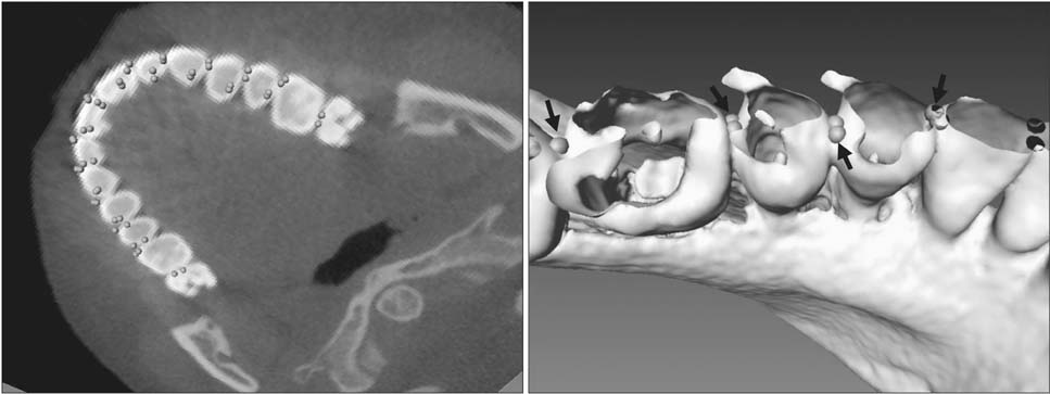
Figure 3 Landmarks represented by arrows on a cone-beam computed tomography (CBCT) slice (left) and a digitally bisected three-dimensional model obtained from the CBCT scans (right) using Avizo software (standard edition version 6.0; Mercury Computer System Inc., Chelmsford, MA, USA).
Cited by 1 articles
-
Reference points suitable for evaluation of the additional arch length required for leveling the curve of Spee
Yong-Hwa Cho, Sung-Hoon Lim, Sung-Nam Gang
Korean J Orthod. 2016;46(6):356-363. doi: 10.4041/kjod.2016.46.6.356.
Reference
-
1. Wedrychowska-Szulc B, Janiszewska-Olszowska J, Stepień P. Overall and anterior Bolton ratio in Class I, II, and III orthodontic patients. Eur J Orthod. 2010; 32:313–318.
Article2. Bolton WA. Disharmony in tooth size and its relation to the analysis and treatment of malocclusion. Angle Orthod. 1958; 28:113–130.3. Bolton WA. The clinical use of a tooth size analysis. Am J Orthod Dentofacial Orthop. 1962; 48:504–529.4. Othman SA, Harradine NW. Tooth-size discrepancy and Bolton's ratios: a literature review. J Orthod. 2006; 33:45–51.
Article5. Sousa MV, Vasconcelos EC, Janson G, Garib D, Pinzan A. Accuracy and reproducibility of 3-dimensional digital model measurements. Am J Orthod Dentofacial Orthop. 2012; 142:269–273.
Article6. Quimby ML, Vig KW, Rashid RG, Firestone AR. The accuracy and reliability of measurements made on computer-based digital models. Angle Orthod. 2004; 74:298–303.7. Mah JK, Huang JC, Choo H. Practical applications of cone-beam computed tomography in orthodontics. J Am Dent Assoc. 2010; 141:Suppl 3. 7S–13S.
Article8. Lagravère MO, Carey J, Toogood RW, Major PW. Three-dimensional accuracy of measurements made with software on cone-beam computed tomography images. Am J Orthod Dentofacial Orthop. 2008; 134:112–116.
Article9. Akyalcin S, Dyer DJ, English JD, Sar C. Comparison of 3-dimensional dental models from different sources: diagnostic accuracy and surface registration analysis. Am J Orthod Dentofacial Orthop. 2013; 144:831–837.
Article10. Flügge TV, Schlager S, Nelson K, Nahles S, Metzger MC. Precision of intraoral digital dental impressions with iTero and extraoral digitization with the iTero and a model scanner. Am J Orthod Dentofacial Orthop. 2013; 144:471–478.
Article11. El-Zanaty HM, El-Beialy AR, Abou El-Ezz AM, Attia KH, El-Bialy AR, Mostafa YA. Three-dimensional dental measurements: An alternative to plaster models. Am J Orthod Dentofacial Orthop. 2010; 137:259–265.
Article12. Kim J, Heo G, Lagravère MO. Accuracy of laserscanned models compared to plaster models and cone-beam computed tomography. Angle Orthod. 2014; 84:443–450.
Article13. Whetten JL, Williamson PC, Heo G, Varnhagen C, Major PW. Variations in orthodontic treatment planning decisions of Class II patients between virtual 3-dimensional models and traditional plaster study models. Am J Orthod Dentofacial Orthop. 2006; 130:485–491.
Article14. Fleming PS, Marinho V, Johal A. Orthodontic measurements on digital study models compared with plaster models: a systematic review. Orthod Craniofac Res. 2011; 14:1–16.
Article15. Naidu D, Freer TJ. Validity, reliability, and reproducibility of the iOC intraoral scanner: a comparison of tooth widths and Bolton ratios. Am J Orthod Dentofacial Orthop. 2013; 144:304–310.
Article16. Wiranto MG, Engelbrecht WP, Tutein Nolthenius HE, van der Meer WJ, Ren Y. Validity, reliability, and reproducibility of linear measurements on digital models obtained from intraoral and cone-beam computed tomography scans of alginate impressions. Am J Orthod Dentofacial Orthop. 2013; 143:140–147.
Article17. Proffit WR. Contemporary orthodontics. 3rd ed. St. Louis: Mosby;2000. p. 170.18. Roberts CT, Richmond S. The design and analysis of reliability studies for the use of epidemiological and audit indices in orthodontics. Br J Orthod. 1997; 24:139–147.
Article19. Shellhart WC, Lange DW, Kluemper GT, Hicks EP, Kaplan AL. Reliability of the Bolton tooth-size analysis when applied to crowded dentitions. Angle Orthod. 1995; 65:327–334.20. Zilberman O, Huggare JA, Parikakis KA. Evaluation of the validity of tooth size and arch width measurements using conventional and three-dimensional virtual orthodontic models. Angle Orthod. 2003; 73:301–306.21. Nalcaci R, Topcuoglu T, Ozturk F. Comparison of Bolton analysis and tooth size measurements obtained using conventional and three-dimensional orthodontic models. Eur J Dent. 2013; 7:Suppl 1. S66–S70.
Article22. Kau CH, Littlefield J, Rainy N, Nguyen JT, Creed B. Evaluation of CBCT digital models and traditional models using the Little's Index. Angle Orthod. 2010; 80:435–439.
Article
- Full Text Links
-
- Actions
-
Cited
- CITED
-
- Close
- Share
-
- Similar articles
-
- Assessment of the accuracy of laser-scanned models and 3-dimensional rendered cone-beam computed tomographic images compared to digital caliper measurements on plaster casts
- Comparison of model analysis measurements among plaster model, laser scan digital model, and cone beam CT image
- Three-dimensional comparison of 2 digital models obtained from cone-beam computed tomographic scans of polyvinyl siloxane impressions and plaster models
- Accuracy and reliability of measurements performed using two different software programs on digital models generated using laser and computed tomography plaster model scanners
- Clinical Validity of Tooth Size Measurements Obtained via Digital Methods with Intraoral Scanning
