Accuracy and reliability of measurements performed using two different software programs on digital models generated using laser and computed tomography plaster model scanners
- Affiliations
-
- 1Department of Orthodontics, School of Dentistry, Universidade Federal Fluminense, Niterói, Brazil. leocamardella@globo.com
- 2Department of Orthodontics and Craniofacial Biology, Radboud University Medical Center, Nijmegen, The Netherlands.
- KMID: 2468637
- DOI: http://doi.org/10.4041/kjod.2020.50.1.13
Abstract
OBJECTIVE
The aim of this study was to compare the accuracy and reliability of measurements performed using two different software programs on digital models generated using two types of plaster model scanners (a laser scanner and a computed tomography [CT] scanner).
METHODS
Thirty plaster models were scanned with a 3Shape laser scanner and with a Flash CT scanner. Two examiners performed measurements on plaster models by using digital calipers and on digital models by using Ortho Analyzer (3Shape) and Digimodel® (OrthoProof) software programs. Forty-two measurements, including tooth diameter, crown height, overjet, overbite, intercanine and intermolar distances, and sagittal relationship, were obtained.
RESULTS
Statistically significant differences were not found between the plaster and digital model measurements (ANOVA); however, some discrepancies were clinically relevant. Plaster and digital model measurements made using the two scanning methods showed high intraclass coefficient correlation values and acceptable 95% limits of agreement in the Bland-Altman analysis. The software used did not influence the accuracy of measurements.
CONCLUSIONS
Digital models generated from plaster casts by using laser and CT scanning and measured using two different software programs are accurate, and the measurements are reliable. Therefore, both fabrication methods and software could be used interchangeably.
Keyword
Figure
-
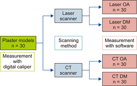
Figure 1 Schematic overview of the design of the study. Laser OA, Digital model produced by laser scanning and measured with the Ortho Analyzer software; Laser DM, digital model produced by laser scanning and measured with the Digimodel software; CT OA, digital model produced by computed tomography (CT) scanning and measured with the Ortho Analyzer software; CT DM, digital model produced by CT scanning and measured with the Digimodel software.
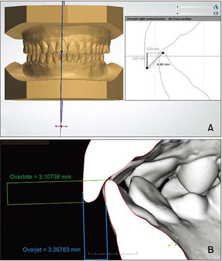
Figure 2 A, Measuring the overbite by using the Ortho Analyzer software. B, Measuring the overjet and overbite by using the Digimodel software.
Cited by 1 articles
-
Evaluation of accuracy of 3-dimensional printed dental models in reproducing intermaxillary relational measurements: Based on inter-operator differences
Won-joon Choi, Su-jung Lee, Cheol-Hyun Moon
Korean J Orthod. 2022;52(1):20-28. doi: 10.4041/kjod.2022.52.1.20.
Reference
-
1. Rischen RJ, Breuning KH, Bronkhorst EM, Kuijpers-Jagtman AM. Records needed for orthodontic diagnosis and treatment planning: a systematic review. PLoS One. 2013; 8:e74186.
Article2. Abizadeh N, Moles DR, O'Neill J, Noar JH. Digital versus plaster study models: how accurate and reproducible are they? J Orthod. 2012; 39:151–159.
Article3. de Waard O, Rangel FA, Fudalej PS, Bronkhorst EM, Kuijpers-Jagtman AM, Breuning KH. Reproducibility and accuracy of linear measurements on dental models derived from cone-beam computed tomography compared with digital dental casts. Am J Orthod Dentofacial Orthop. 2014; 146:328–336.
Article4. Torassian G, Kau CH, English JD, Powers J, Bussa HI, Marie Salas-Lopez A, et al. Digital models vs plaster models using alginate and alginate substitute materials. Angle Orthod. 2010; 80:474–481.
Article5. White AJ, Fallis DW, Vandewalle KS. Analysis of intra-arch and interarch measurements from digital models with 2 impression materials and a modeling process based on cone-beam computed tomography. Am J Orthod Dentofacial Orthop. 2010; 137:456.e1–456.e9. discussion 456–7.
Article6. Ahn HW, Chang YJ, Kim KA, Joo SH, Park YG, Park KH. Measurement of three-dimensional perioral soft tissue changes in dentoalveolar protrusion patients after orthodontic treatment using a structured light scanner. Angle Orthod. 2014; 84:795–802.
Article7. Grünheid T, Patel N, De Felippe NL, Wey A, Gaillard PR, Larson BE. Accuracy, reproducibility, and time efficiency of dental measurements using different technologies. Am J Orthod Dentofacial Orthop. 2014; 145:157–164.
Article8. Asquith J, Gillgrass T, Mossey P. Three-dimensional imaging of orthodontic models: a pilot study. Eur J Orthod. 2007; 29:517–522.
Article9. Stevens DR, Flores-Mir C, Nebbe B, Raboud DW, Heo G, Major PW. Validity, reliability, and reproducibility of plaster vs digital study models: comparison of peer assessment rating and Bolton analysis and their constituent measurements. Am J Orthod Dentofacial Orthop. 2006; 129:794–803.
Article10. Mullen SR, Martin CA, Ngan P, Gladwin M. Accuracy of space analysis with emodels and plaster models. Am J Orthod Dentofacial Orthop. 2007; 132:346–352.
Article11. Horton HM, Miller JR, Gaillard PR, Larson BE. Technique comparison for efficient orthodontic tooth measurements using digital models. Angle Orthod. 2010; 80:254–261.
Article12. Goonewardene RW, Goonewardene MS, Razza JM, Murray K. Accuracy and validity of space analysis and irregularity index measurements using digital models. Aust Orthod J. 2008; 24:83–90.13. Sousa MV, Vasconcelos EC, Janson G, Garib D, Pinzan A. Accuracy and reproducibility of 3-dimensional digital model measurements. Am J Orthod Dentofacial Orthop. 2012; 142:269–273.
Article14. Costalos PA, Sarraf K, Cangialosi TJ, Efstratiadis S. Evaluation of the accuracy of digital model analysis for the American Board of Orthodontics objective grading system for dental casts. Am J Orthod Dentofacial Orthop. 2005; 128:624–629.
Article15. Keating AP, Knox J, Bibb R, Zhurov AI. A comparison of plaster, digital and reconstructed study model accuracy. J Orthod. 2008; 35:191–201. discussion 175.
Article16. Kim J, Heo G, Lagravère MO. Accuracy of laser-scanned models compared to plaster models and cone-beam computed tomography. Angle Orthod. 2014; 84:443–450.
Article17. Okunami TR, Kusnoto B, BeGole E, Evans CA, Sadowsky C, Fadavi S. Assessing the American Board of Orthodontics objective grading system: digital vs plaster dental casts. Am J Orthod Dentofacial Orthop. 2007; 131:51–56.
Article18. Bootvong K, Liu Z, McGrath C, Hägg U, Wong RW, Bendeus M, et al. Virtual model analysis as an alternative approach to plaster model analysis: reliability and validity. Eur J Orthod. 2010; 32:589–595.
Article19. Tomassetti JJ, Taloumis LJ, Denny JM, Fischer JR Jr. A comparison of 3 computerized Bolton tooth-size analyses with a commonly used method. Angle Orthod. 2001; 71:351–357.20. Hayashi K, Sachdeva AU, Saitoh S, Lee SP, Kubota T, Mizoguchi I. Assessment of the accuracy and reliability of new 3-dimensional scanning devices. Am J Orthod Dentofacial Orthop. 2013; 144:619–625.
Article21. Creed B, Kau CH, English JD, Xia JJ, Lee RP. A comparison of the accuracy of linear measurements obtained from cone beam computerized tomography images and digital models. Semin Orthod. 2011; 17:49–56.
Article22. Hildebrand JC, Palomo JM, Palomo L, Sivik M, Hans M. Evaluation of a software program for applying the American Board of Orthodontics objective grading system to digital casts. Am J Orthod Dentofacial Orthop. 2008; 133:283–289.
Article23. Santoro M, Galkin S, Teredesai M, Nicolay OF, Cangialosi TJ. Comparison of measurements made on digital and plaster models. Am J Orthod Dentofacial Orthop. 2003; 124:101–105.
Article24. De Luca Canto G, Pachêco-Pereira C, Lagravere MO, Flores-Mir C, Major PW. Intra-arch dimensional measurement validity of laser-scanned digital dental models compared with the original plaster models: a systematic review. Orthod Craniofac Res. 2015; 18:65–76.
Article25. Watanabe-Kanno GA, Abrão J, Miasiro Junior H, Sánchez-Ayala A, Lagravère MO. Reproducibility, reliability and validity of measurements obtained from Cecile3 digital models. Braz Oral Res. 2009; 23:288–295.
Article26. Veenema AC, Katsaros C, Boxum SC, Bronkhorst EM, Kuijpers-Jagtman AM. Index of complexity, outcome and need scored on plaster and digital models. Eur J Orthod. 2009; 31:281–286.
Article27. Wan Hassan WN, Othman SA, Chan CS, Ahmad R, Ali SN, Abd Rohim A. Assessing agreement in measurements of orthodontic study models: digital caliper on plaster models vs 3-dimensional software on models scanned by structured-light scanner. Am J Orthod Dentofacial Orthop. 2016; 150:886–895.
Article28. Westerlund A, Tancredi W, Ransjö M, Bresin A, Psonis S, Torgersson O. Digital casts in orthodontics: a comparison of 4 software systems. Am J Orthod Dentofacial Orthop. 2015; 147:509–516.
Article29. Pandis N. Sample calculations for comparison of 2 means. Am J Orthod Dentofacial Orthop. 2012; 141:519–521.
Article30. Naidu D, Freer TJ. Validity, reliability, and reproducibility of the iOC intraoral scanner: a comparison of tooth widths and Bolton ratios. Am J Orthod Dentofacial Orthop. 2013; 144:304–310.
Article31. Fleming PS, Marinho V, Johal A. Orthodontic measurements on digital study models compared with plaster models: a systematic review. Orthod Craniofac Res. 2011; 14:1–16.
Article32. Wiranto MG, Engelbrecht WP, Tutein Nolthenius HE, van der Meer WJ, Ren Y. Validity, reliability, and reproducibility of linear measurements on digital models obtained from intraoral and cone-beam computed tomography scans of alginate impressions. Am J Orthod Dentofacial Orthop. 2013; 143:140–147.
Article
- Full Text Links
-
- Actions
-
Cited
- CITED
-
- Close
- Share
-
- Similar articles
-
- Accuracy of Bolton analysis measured in laser scanned digital models compared with plaster models (gold standard) and cone-beam computer tomography images
- Assessment of the accuracy of laser-scanned models and 3-dimensional rendered cone-beam computed tomographic images compared to digital caliper measurements on plaster casts
- Comparison of model analysis measurements among plaster model, laser scan digital model, and cone beam CT image
- Evaluation of accuracy of 3-dimensional printed dental models in reproducing intermaxillary relational measurements: Based on inter-operator differences
- Clinical Validity of Tooth Size Measurements Obtained via Digital Methods with Intraoral Scanning
