Pneumopericardium: A Rare Complication of Pericardiocentesis
- Affiliations
-
- 1Department of Cardiology, Clinical Echocardiography Laboratory, Intercommunity Hospital of Southern Alps, Gap, France. slamaiskander@live.fr
- KMID: 2160996
- DOI: http://doi.org/10.4250/jcu.2016.24.1.55
Abstract
- Pneumopericardium is defined by the presence of air in the pericardial cavity. It is a rare entity occurring most commonly after trauma. Pneumopericardium resulting after pericardiocentesis is even rarer. We report a case of 46-year-old man, with end-stage renal disease on chronic hemodialysis and who developed a large circumferential pericardial effusion of 40 mm in diastole with swinging heart and diastolic right atrium collapse requiring pericardiocentesis. Few days after, the patient complained of pleuritic chest pain and echocardiogram revealed several tiny sparkling echogenic spots swirling in the pericardial sac. Computed tomography scans revealed a marked anterior pneumopericardium that was conservatively managed.
MeSH Terms
Figure
-
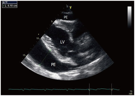
Fig. 1 Two dimensional echocardiography. Parasternal long-axis view showing a large circumferential pericardial effusion of 40 mm in diastole. LV: left ventricle, PE: pericardial effusion.
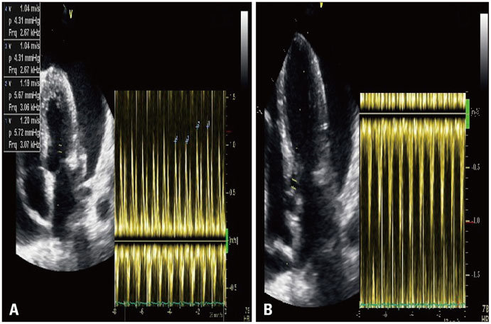
Fig. 2 Pulsed wave Doppler mode showing no significant respiratory variations of the mitral inflow (A) and aortic flow (B).
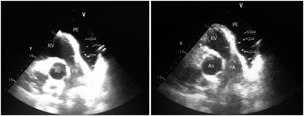
Fig. 3 Two dimensional echocardiography. Parasternal short-axis view showing several tiny sparkling echogenic spots swirling in the pericardial sac evoking micro air bubbles (open arrows). PE: pericardial effusion, RV: right ventricle, Ao: aorta, PA: pulmonary artery.
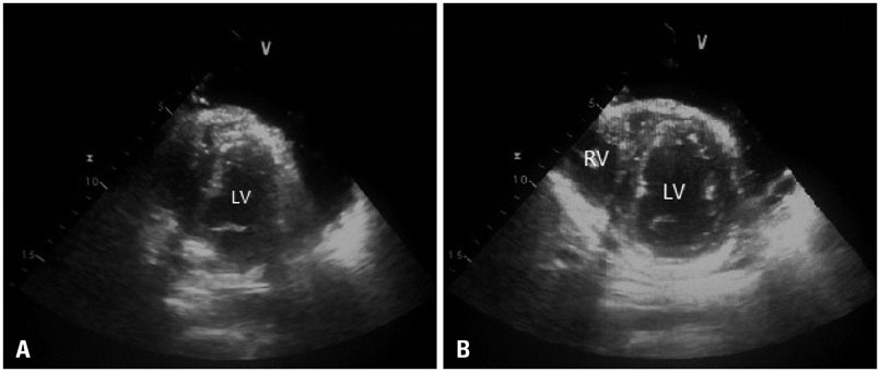
Fig. 4 Two dimensional echocardiography. Parasternal short-axis view (mid-ventricular) showing a partial disappearance of the shape of cardiac chambers in late systole with a band of echoes within (A). Reappearance of cardiac cavities in late diastole (B). RV: right ventricle, LV: left ventricle.
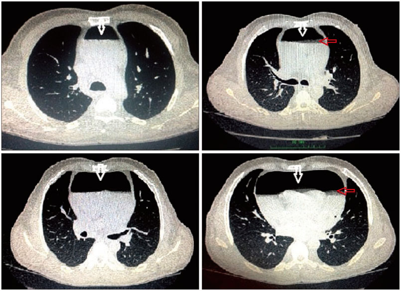
Fig. 5 Chest computed tomography scan revealed a marked pneumopericardium with an anterior extent (white arrows) associated to small amount of pericardial fluid signing a hemopericard (dark red arrows).
Reference
-
1. Lee YJ, Jin SW, Jang SH, Jang YS, Lee EK, Kim YJ, Lee MY, Park JC, Rho TH, Kim JH, Hong SJ, Choi KB. A case of spontaneous pneumomediastinum and pneumopericardium in a young adult. Korean J Intern Med. 2001; 16:205–209.2. Mullens W, Dupont M, De Raedt H. Pneumopericardium after pericardiocentesis. Int J Cardiol. 2007; 118:e57.3. Choi WH, Hwang YM, Park MY, Lee SJ, Lee HY, Kim SW, Jun BY, Min JS, Shin WS, Lee JM, Koh YS, Jeon HK, Chung WS, Seung KB. Pneumopericardium as a complication of pericardiocentesis. Korean Circ J. 2011; 41:280–282.4. Brander L, Ramsay D, Dreier D, Peter M, Graeni R. Continuous left hemidiaphragm sign revisited: a case of spontaneous pneumopericardium and literature review. Heart. 2002; 88:e5.5. Yuce M, Sari I, Davutoglu V, Ozer O, Usalan C. Bubbles around the heart: pneumopericardium 10 days after pericardiocentesis. Echocardiography. 2010; 27:E115–E116.6. Lee SH, Kim WH, Lee SR, Rhee KS, Chae JK, Ko JK. Cardiac tamponade by iatrogenic pneumopericardium. J Cardiovasc Ultrasound. 2008; 16:26–28.7. Rashid MA, Wikström T, Ortenwall P. Pneumopericardium and pneumoperitoneum after penetrating chest injury. Eur J Surg. 1999; 165:278–279.8. Müller AM, Betz MJ, Kromeier J, Ghanem NA, Geibel A, Imdahl A, Frydrychowicz AP. Images in cardiovascular medicine. Acute pneumopericardium due to intestino-pericardial fistula. Circulation. 2006; 114:e7–e9.9. Shackelford RT. Hydropneumopericardium: report of a case with a summary of the literature. JAMA. 1931; 96:187–191.10. Cummings RG, Wesly RL, Adams DH, Lowe JE. Pneumopericardium resulting in cardiac tamponade. Ann Thorac Surg. 1984; 37:511–518.11. Marijon E, Jani D, Aubert S. Unusual chest radiograph after surgical pericardiotomy. Tex Heart Inst J. 2007; 34:256–257.12. Bejvan SM, Godwin JD. Pneumomediastinum: old signs and new signs. AJR Am J Roentgenol. 1996; 166:1041–1048.13. Reid CL, Chandraratna AN, Kawanishi D, Bezdek WD, Schatz R, Nanna M, Rahimtoola SH. Echocardiographic detection of pneumomediastinum and pneumopericardium: the air gap sign. J Am Coll Cardiol. 1983; 1:916–921.14. Kerut EK, Hannawalt C, Everson CT, Nanda NC. The air gap sign. Echocardiography. 2014; 31:400–401.15. Antonini-Canterin F, Nicolosi GL, Mascitelli L, Zanuttini D. Direct demonstration of an air-fluid interface by two-dimensional echocardiography: a new diagnostic sign of hydropneumopericardium. J Am Soc Echocardiogr. 1996; 9:187–189.16. Uluçam MZ. An extremely rare combination: pneumopericardium, pneumoperitoneum, and subcutanous emphysema-a case report. Cardiol Ther. 2013; 2:103–110.17. Ozerkan F, Bilgin M, Oktem MS, Alkan MB. [Pneumopericardium after pericardiocentesis: a case report]. Turk Kardiyol Dern Ars. 2011; 39:697–700.18. Wacker W, Merrill JP. Uremic pericarditis in acute and chronic renal failure. J Am Med Assoc. 1954; 156:764–765.19. Rostand SG, Rutsky EA. Pericarditis in end-stage renal disease. Cardiol Clin. 1990; 8:701–707.
- Full Text Links
-
- Actions
-
Cited
- CITED
-
- Close
- Share
-
- Similar articles
-
- Cardiac Tamponade by Iatrogenic Pneumopericardium
- Pneumopericardium as a Complication of Pericardiocentesis
- Tension Pneumopericardium after Pericardiocentesis
- A Case of Spontaneous Pneumomediastinum and Pneumopericardium in a Patient with Acute Exacerbation of Idiopathic Pulmonary Fibrosis
- Negative Pressure Pulmonary Edema Together with Pneumopericardium after General Anesthesia
