Cardiac Tamponade by Iatrogenic Pneumopericardium
- Affiliations
-
- 1Division of Cardiology, Department of Internal Medicine, Chonbuk National University Medical School, Jeonju, Korea. jkko@chonbuk.ac.kr
- KMID: 1486607
- DOI: http://doi.org/10.4250/jcu.2008.16.1.26
Abstract
- Pneumopericardium is defined as the condition of presence of air in the pericardial space. It is associated with various etiologies such as chest trauma, infection or invasive procedures. We herein describe a case of cardiac tamponade associated with pneumopericardium. We diagnosed iatrogenic pneumopericardium by plain chest radiography and two-dimensional echocardiography. The patient was successfully treated by re-pericardiocentesis.
Figure
-
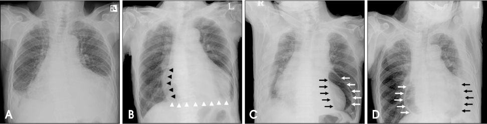
Fig. 1 A: Chest X-ray showed cardiomegaly with bilteral pleural effusions at admission. B: Chest PA on third hospital day demonstrated cardiomegaly with the line separating pericardium from the heart (black arrow heads) and "continuous diaphragm sign" (white arrow heads) implying presence of air in the pericardial cavity, pneumopericardium. C-D: Both decubitus views revealed thin parietal pericardium (white arrows) and air-fluid level (black arrows) in the pericardial cavity, which suggests hydropneumopericardium.

Fig. 2 Two-dimensional echocardiography [A: apical four-chamber view, B: parasternal short axis view, papillary muscle level] showed pericardial effusion (PE) with the "swirling bubbles sign" that indicated multiple unusual tiny echo-bright spots (white arrows) in pericardial cavity, suggesting formation of air microbubbles through continuous shaking of the air-fluid surface with heart beats. LV: left ventricle, LA: left atrium, RA: right atrium.

Fig. 3 Cine-fluoroscopy demonstrated large multiple lumps of air (black arrows) moving in the pericardial cavity with the heart beats.
Reference
-
1. Guberman BA, Fowler NO, Engel PJ, Gueron M, Allen JM. Cardiac tamponade in medical patients. Circulation. 1981. 64:633–640.
Article2. Spodick DH. Acute cardiac tamponade. N Engl J Med. 2003. 349:684–690.
Article3. Han SW, Kim KS, Hur SH, Hyun DW, Kim YN, Kim KB. Efficacy of 2-dimensional contrast echocardiography during pericardiocentesis. J Kor Soc Echocardiogr. 1996. 4:80–84.
Article4. Tsang TS, Enriquez-Sarano M, Freeman WK, Barnes ME, Sinak LJ, Gersh BJ, Bailey KR, Seward JB. Consecutive 1127 therapeutic echocardiographically guided pericardiocentesis: clinical profile, practice patterns, and outcomes spanning 21 years. Mayo Clin Proc. 2002. 77:429–436.
Article5. Krikorian JG, Hancock EW. Pericardiocentesis. Am J Med. 1978. 65:808–814.
Article6. Bernal JM, Pradhan J, Li T, Tchokonte R, Afonso L. Acute pulmonary edema following pericardiocentesis for cardiac tamponade. Can J Cardiol. 2007. 23:1155–1156.
Article7. Bhindi R, Rees DM. Pneumopericardium masquerading as an acute myocardial infarction. Int J Cardiol. 2007. 114:e83–e84.
Article8. Albrecht CA, Jafri A, Linville L, Anderson V. Cocaine-induced pneumopericardium. Circulation. 2000. 102:2792–2794.
Article9. Rashid MA, Wikström T, Örtenwall P. Pneumopericardium and pneumoperitoneum after penetrating chest injury. Eur J Surg. 1999. 165:278–279.
Article10. Brander L, Ramsay D, Dreier D, Peter M, Graeni R. Continuous left hemodiaphragm sign revisited: a case of spontaneous pneumopericardium and literature review. Heart. 2002. 88:e5.11. Müller AMS, Betz MJ, Kromeier J, Ghanem NA, Geibel A, Imdahl A, Frydrychowicz AP. Acute pneumopericardium due to intestine-pericardial fistula. Circulation. 2006. 114:e7–e9.12. van Haren FMP, Borstlap A, Fouraine N. Tension pneumopericardium in Hodgkin's diasease. Circulation. 2006. 114:e77–e79.13. Reid CL, Chandraratna AN, Kawanishi D, Bezdek WD, Schatz R, Nanna M, Rahimtoola SH. Echocardiographic detection of pneumomediastinum and pneumopericardium: the air gap sign. J Am Coll Cardiol. 1983. 1:916–921.
Article14. Bedotto JB, McBride W, Abraham M, Taylor AL. Echocardiographic diagnosis of pneumopericardium and hydropneumopericardium. J Am Soc Echocardiogr. 1988. 1:359–361.
Article15. Antonini-Canterin F, Nicolosi GL, Mascitelli L, Zanuttini D. Direct demonstration of an air-fluid interface by two-dimensional echocardiography: a new diagnostic sign of hydropneumopericardium. J Am Soc Echocardiogr. 1996. 9:187–189.
Article16. Mullens W, Dupont M, De Raedt H. Pneumopericardium after pericardiocentesis. Int J Cardiol. 2007. 118:e57.
Article
- Full Text Links
-
- Actions
-
Cited
- CITED
-
- Close
- Share
-
- Similar articles
-
- Tension Pneumopericardium after Pericardiocentesis
- Neonatal Tension Pneumopericardium
- Pneumopericardium as a Complication of Pericardiocentesis
- Successful Treatment of Cardiac Tamponade Associated with Umbilical Venous Catheter by Pericardiocentesis in Two Neonates: Two Case Reports
- A successfully treated case of primary purulent pericarditis complicated by cardiac tamponade and pneumopericardium
