Korean J Radiol.
2015 Oct;16(5):1153-1162. 10.3348/kjr.2015.16.5.1153.
Relationship between Myocardial Extracellular Space Expansion Estimated with Post-Contrast T1 Mapping MRI and Left Ventricular Remodeling and Neurohormonal Activation in Patients with Dilated Cardiomyopathy
- Affiliations
-
- 1Cardiology Division, Department of Internal Medicine, Yonsei University College of Medicine, Seoul 06273, Korea. CHOI0928@yuhs.ac
- 2Department of Radiology, Yonsei University College of Medicine, Seoul 06273, Korea.
- KMID: 2160783
- DOI: http://doi.org/10.3348/kjr.2015.16.5.1153
Abstract
OBJECTIVE
Post-contrast T1 values are closely related to the degree of myocardial extracellular space expansion. We determined the relationship between post-contrast T1 values and left ventricular (LV) diastolic function, LV remodeling, and neurohormonal activation in patients with dilated cardiomyopathy (DCM).
MATERIALS AND METHODS
Fifty-nine patients with DCM (mean age, 55 +/- 15 years; 41 males and 18 females) who underwent both 1.5T magnetic resonance imaging and echocardiography were enrolled. The post-contrast 10-minute T1 value was generated from inversion time scout images obtained using the Look-Locker inversion recovery sequence and a curve-fitting algorithm. The T1 sample volume was obtained from three interventricular septal points, and the mean T1 value was used for analysis. The N-Terminal pro-B-type natriuretic peptide (NT-proBNP) level was measured in 40 patients.
RESULTS
The mean LV ejection fraction was 24 +/- 9% and the post-T1 value was 254.5 +/- 46.4 ms. The post-contrast T1 value was significantly correlated with systolic longitudinal septal velocity (s'), peak late diastolic velocity of the mitral annulus (a'), the diastolic elastance index (Ed, [E/e']/stroke volume), LV mass/volume ratio, LV end-diastolic wall stress, and LV end-systolic wall stress. In a multivariate analysis without NT-proBNP, T1 values were independently correlated with Ed (beta = -0.351, p = 0.016) and the LV mass/volume ratio (beta = 0.495, p = 0.001). When NT-proBNP was used in the analysis, NT-proBNP was independently correlated with the T1 values (beta = -0.339, p = 0.017).
CONCLUSION
Post-contrast T1 is closely related to LV remodeling, diastolic function, and neurohormonal activation in patients with DCM.
MeSH Terms
-
Adult
Aged
Algorithms
Blood Pressure
Cardiomyopathy, Dilated/*diagnosis/radiography
Echocardiography
Extracellular Space/physiology/*radiography
Female
Heart Ventricles/physiopathology/radiography
Humans
Magnetic Resonance Imaging
Male
Middle Aged
Multivariate Analysis
Natriuretic Peptide, Brain/analysis
Peptide Fragments/analysis
*Ventricular Remodeling
Natriuretic Peptide, Brain
Peptide Fragments
Figure
-
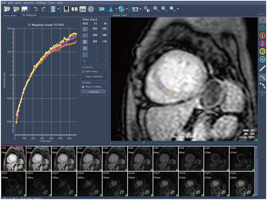
Fig. 1 Representative case of measuring post-T1 value in Look-Locker-based inversion time scout images.
Reference
-
1. Jessup M, Brozena S. Heart failure. N Engl J Med. 2003; 348:2007–2018.2. Elliott P, Andersson B, Arbustini E, Bilinska Z, Cecchi F, Charron P, et al. Classification of the cardiomyopathies: a position statement from the European Society Of Cardiology Working Group on Myocardial and Pericardial Diseases. Eur Heart J. 2008; 29:270–276.3. Roger VL, Weston SA, Redfield MM, Hellermann-Homan JP, Killian J, Yawn BP, et al. Trends in heart failure incidence and survival in a community-based population. JAMA. 2004; 292:344–350.4. Rihal CS, Nishimura RA, Hatle LK, Bailey KR, Tajik AJ. Systolic and diastolic dysfunction in patients with clinical diagnosis of dilated cardiomyopathy. Relation to symptoms and prognosis. Circulation. 1994; 90:2772–2779.5. Pinamonti B, Zecchin M, Di Lenarda A, Gregori D, Sinagra G, Camerini F. Persistence of restrictive left ventricular filling pattern in dilated cardiomyopathy: an ominous prognostic sign. J Am Coll Cardiol. 1997; 29:604–612.6. Doust JA, Pietrzak E, Dobson A, Glasziou P. How well does B-type natriuretic peptide predict death and cardiac events in patients with heart failure: systematic review. BMJ. 2005; 330:625.7. Kwon DH, Halley CM, Carrigan TP, Zysek V, Popovic ZB, Setser R, et al. Extent of left ventricular scar predicts outcomes in ischemic cardiomyopathy patients with significantly reduced systolic function: a delayed hyperenhancement cardiac magnetic resonance study. JACC Cardiovasc Imaging. 2009; 2:34–44.8. Rudolph A, Abdel-Aty H, Bohl S, Boyé P, Zagrosek A, Dietz R, et al. Noninvasive detection of fibrosis applying contrast-enhanced cardiac magnetic resonance in different forms of left ventricular hypertrophy relation to remodeling. J Am Coll Cardiol. 2009; 53:284–291.9. Iles L, Pfluger H, Phrommintikul A, Cherayath J, Aksit P, Gupta SN, et al. Evaluation of diffuse myocardial fibrosis in heart failure with cardiac magnetic resonance contrast-enhanced T1 mapping. J Am Coll Cardiol. 2008; 52:1574–1580.10. Karaahmet T, Tigen K, Dundar C, Pala S, Guler A, Kilicgedik A, et al. The effect of cardiac fibrosis on left ventricular remodeling, diastolic function, and N-terminal pro-B-type natriuretic peptide levels in patients with nonischemic dilated cardiomyopathy. Echocardiography. 2010; 27:954–960.11. Mewton N, Liu CY, Croisille P, Bluemke D, Lima JA. Assessment of myocardial fibrosis with cardiovascular magnetic resonance. J Am Coll Cardiol. 2011; 57:891–903.12. Lang RM, Bierig M, Devereux RB, Flachskampf FA, Foster E, Pellikka PA, et al. Recommendations for chamber quantification: a report from the American Society of Echocardiography's Guidelines and Standards Committee and the Chamber Quantification Writing Group, developed in conjunction with the European Association of Echocardiography, a branch of the European Society of Cardiology. J Am Soc Echocardiogr. 2005; 18:1440–1463.13. Severino S, Caso P, Galderisi M, De Simone L, Petrocelli A, de Divitiis O, et al. Use of pulsed Doppler tissue imaging to assess regional left ventricular diastolic dysfunction in hypertrophic cardiomyopathy. Am J Cardiol. 1998; 82:1394–1398.14. Quiñones MA, Otto CM, Stoddard M, Waggoner A, Zoghbi WA;. Recommendations for quantification of Doppler echocardiography: a report from the Doppler Quantification Task Force of the Nomenclature and Standards Committee of the American Society of Echocardiography. J Am Soc Echocardiogr. 2002; 15:167–184.15. Nagueh SF, Middleton KJ, Kopelen HA, Zoghbi WA, Quiñones MA. Doppler tissue imaging: a noninvasive technique for evaluation of left ventricular relaxation and estimation of filling pressures. J Am Coll Cardiol. 1997; 30:1527–1533.16. Redfield MM, Jacobsen SJ, Borlaug BA, Rodeheffer RJ, Kass DA. Age- and gender-related ventricular-vascular stiffening: a community-based study. Circulation. 2005; 112:2254–2262.17. Kelly RP, Ting CT, Yang TM, Liu CP, Maughan WL, Chang MS, et al. Effective arterial elastance as index of arterial vascular load in humans. Circulation. 1992; 86:513–521.18. McCrohon JA, Moon JC, Prasad SK, McKenna WJ, Lorenz CH, Coats AJ, et al. Differentiation of heart failure related to dilated cardiomyopathy and coronary artery disease using gadolinium-enhanced cardiovascular magnetic resonance. Circulation. 2003; 108:54–59.19. Park S, Choi BW, Rim SJ, Shim CY, Ko YG, Kang SM, et al. Delayed hyperenhancement magnetic resonance imaging is useful in predicting functional recovery of nonischemic left ventricular systolic dysfunction. J Card Fail. 2006; 12:93–99.20. Amano Y, Takayama M, Kumita S. Contrast-enhanced myocardial T1-weighted scout (Look-Locker) imaging for the detection of myocardial damages in hypertrophic cardiomyopathy. J Magn Reson Imaging. 2009; 30:778–784.21. Karlsson M, Nordell B. Analysis of the Look-Locker T(1) mapping sequence in dynamic contrast uptake studies: simulation and in vivo validation. Magn Reson Imaging. 2000; 18:947–954.22. Alter P, Rupp H, Stoll F, Adams P, Figiel JH, Klose KJ, et al. Increased end diastolic wall stress precedes left ventricular hypertrophy in dilative heart failure--use of the volume-based wall stress index. Int J Cardiol. 2012; 157:233–238.23. White SK, Sado DM, Flett AS, Moon JC. Characterising the myocardial interstitial space: the clinical relevance of non-invasive imaging. Heart. 2012; 98:773–779.24. Nacif MS, Turkbey EB, Gai N, Nazarian S, van der Geest RJ, Noureldin RA, et al. Myocardial T1 mapping with MRI: comparison of look-locker and MOLLI sequences. J Magn Reson Imaging. 2011; 34:1367–1373.25. Ho CY, Abbasi SA, Neilan TG, Shah RV, Chen Y, Heydari B, et al. T1 measurements identify extracellular volume expansion in hypertrophic cardiomyopathy sarcomere mutation carriers with and without left ventricular hypertrophy. Circ Cardiovasc Imaging. 2013; 6:415–422.26. Pinamonti B, Di Lenarda A, Sinagra G, Camerini F. Restrictive left ventricular filling pattern in dilated cardiomyopathy assessed by Doppler echocardiography: clinical, echocardiographic and hemodynamic correlations and prognostic implications. Heart Muscle Disease Study Group. J Am Coll Cardiol. 1993; 22:808–815.27. Choi EY, Choi BW, Kim SA, Rhee SJ, Shim CY, Kim YJ, et al. Patterns of late gadolinium enhancement are associated with ventricular stiffness in patients with advanced non-ischaemic dilated cardiomyopathy. Eur J Heart Fail. 2009; 11:573–580.28. Tham EB, Haykowsky MJ, Chow K, Spavor M, Kaneko S, Khoo NS, et al. Diffuse myocardial fibrosis by T1-mapping in children with subclinical anthracycline cardiotoxicity: relationship to exercise capacity, cumulative dose and remodeling. J Cardiovasc Magn Reson. 2013; 15:48.29. Creemers EE, Pinto YM. Molecular mechanisms that control interstitial fibrosis in the pressure-overloaded heart. Cardiovasc Res. 2011; 89:265–272.30. Tsuruda T, Boerrigter G, Huntley BK, Noser JA, Cataliotti A, Costello-Boerrigter LC, et al. Brain natriuretic Peptide is produced in cardiac fibroblasts and induces matrix metalloproteinases. Circ Res. 2002; 91:1127–1134.
- Full Text Links
-
- Actions
-
Cited
- CITED
-
- Close
- Share
-
- Similar articles
-
- Erratum: Relationship between Myocardial Extracellular Space Expansion Estimated with Post-Contrast T1 Mapping MRI and Left Ventricular Remodeling and Neurohormonal Activation in Patients with Dilated Cardiomyopathy
- Left Ventricular Dysfunction and Dilated Cardiomyopathy in Infants and Children with Wolff-Parkinson-White Syndrome in the Absence of Tachyarrhythmias
- Hemodynamics and Left Ventricular Cineangiographic Findings in Idiopathic Dilated Cardiomyopathy
- Catheter Ablation of Ventricular Tachycardia in Patients with Post-Infarction Cardiomyopathy
- Clinical Application of T1 and T2 Mapping in Cardiac Magnetic Resonance Imaging for Nonischemic Cardiomyopathy: A Case-Based Review
