Role of cone-beam computed tomography in the evaluation of a paradental cyst related to the fusion of a wisdom tooth with a paramolar: A rare case report
- Affiliations
-
- 1Department of Oral and Maxillofacial Radiology, Faculty of Dentistry, Erciyes University, Kayseri, Turkey. gozcan89@gmail.com
- 2Department of Oral and Maxillofacial Surgery, Faculty of Dentistry, Gaziosmanpasa University, Tokat, Turkey.
- 3Department of Pathology, Yozgat State Hospital, Yozgat, Turkey.
- KMID: 2160159
- DOI: http://doi.org/10.5624/isd.2016.46.1.57
Abstract
- Fusion is an abnormality of tooth development defined as the union of two developing dental germs, resulting in a single large dental structure. This irregular tooth morphology is associated with a high predisposition to dental caries and periodontal diseases. As a result of recurring inflammatory periodontal processes, disorders such as periodontal pocket, pericoronitis, and paradental cysts may develop. A rare mandibular anatomic variation is the retromolar canal, which is very significant for surgical procedures. The fusion of a paramolar and mandibular third molar associated with a paradental cyst co-occurring with the presence of a retromolar canal is rare, and the aim of the present study is to describe the evaluation of this anatomical configuration using cone-beam computed tomography.
MeSH Terms
Figure
-
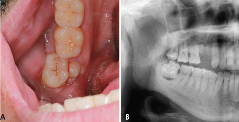
Fig. 1 An intraoral examination shows an extra cusp on the lingual aspect of the third molar tooth and inflamed gingiva around the abnormal tooth formation. B. A cropped panoramic radiograph exhibits an extra tooth in close association with the mandibular third molar and a sharply delineated radiolucent lesion around the apical area in relation to the same molar.
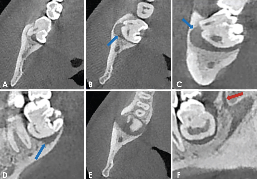
Fig. 2 Cone-beam tomographic images of the right mandibular third molar region demonstrate the fusion of a supernumerary molar on the lingual aspect of the third molar on the axial (A and B), coronal (C), and sagittal (D) planes. B-D. A cystic lesion can be identified around the fused teeth (arrows). E. The axial image shows the lesion in relation to the fused teeth. F. The retromolar canal is observed behind the fused teeth in the sagittal view (arrow).

Fig. 3 The extracted fused teeth. A. Superior aspect of the fused teeth. B. Buccal aspect. C. Lingual aspect. D. inferior aspect.

Fig. 4 A. A photomicrograph shows the cystic lining with mixed dense inflammatory infiltrate (hematoxylin and eosin stain, 40×). B. 200× magnification.
Reference
-
1. Nunes E, de Moraes IG, de Novaes PM, de Sousa SM. Bilateral fusion of mandibular second molars with supernumerary teeth: case report. Braz Dent J. 2002; 13:137–141.
Article2. Neves AA, Neves ML, Farinhas JA. Bilateral connation of permanent mandibular incisors: a case report. Int J Paediatr Dent. 2002; 12:61–65.
Article3. Levitas TC. Gemination, fusion, twinning and concrescence. ASDC J Dent Child. 1965; 32:93–100.4. Muthukumar RS, Arunkumar S, Sadasiva K. Bilateral fusion of mandibular second premolar and supernumerary tooth: a rare case report. J Oral Maxillofac Pathol. 2012; 16:128–130.
Article5. Prakash AR, Reddy PS, Rajanikanth M. Paradental cyst associated with supernumerary tooth fused with third molar: a rare case report. J Oral Maxillofac Pathol. 2012; 16:131–133.
Article6. Mufeed A, Chatra L, Shenai P. Diagnostic features of the paradental cyst and report of a case. Dentomaxillofac Radiol. 2009; 38:125–126.
Article7. Bilecenoglu B, Tuncer N. Clinical and anatomical study of retromolar foramen and canal. J Oral Maxillofac Surg. 2006; 64:1493–1497.
Article8. von Arx T, Hänni A, Sendi P, Buser D, Bornstein MM. Radiographic study of the mandibular retromolar canal: an anatomic structure with clinical importance. J Endod. 2011; 37:1630–1635.
Article9. Rossi AC, Freire AR, Prado GB, Prado FB, Botacin PR, Caria PH. Incidence of retromolar foramen in human mandibles: ethnic and clinical aspects. Int J Morphol. 2012; 30:1074–1078.
Article10. Pires CA, Bissada NF, Becker JJ, Kanawati A, Landers MA. Mandibular incisive canal: cone beam computed tomography. Clin Implant Dent Relat Res. 2012; 14:67–73.
Article11. Watson RM, Davis DM, Forman GH, Coward T. Considerations in design and fabrication of maxillary implant-supported prostheses. Int J Prosthodont. 1991; 4:232–239.12. Neves FS, Souza TC, Almeida SM, Haiter-Neto F, Freitas DQ, Bóscolo FN. Correlation of panoramic radiography and cone beam CT findings in the assessment of the relationship between impacted mandibular third molars and the mandibular canal. Dentomaxillofac Radiol. 2012; 41:553–557.
Article13. Chen HS, Huang YL. Fusion of third and fourth mandibular molars? Oral Surg Oral Med Oral Pathol. 1992; 73:767.
Article14. Järvinen S, Lehtinen L. Supernumerary and congenitally missing primary teeth in Finnish children. An epidemiologic study. Acta Odontol Scand. 1980; 39:83–86.15. Parolia A, Kundabala M, Dahal M, Mohan M, Thomas MS. Management of supernumerary teeth. J Conserv Dent. 2011; 14:221–224.
Article16. Ghoddusi J, Zarei M, Jafarzadeh H. Endodontic treatment of a supernumerary tooth fused to a mandibular second molar: a case report. J Oral Sci. 2006; 48:39–41.
Article17. Indra R, Srinivasan MR, Farzana H, Karthikeyan K. Endodontic management of a fused maxillary lateral incisor with a supernumerary tooth: a case report. J Endod. 2006; 32:1217–1219.
Article18. Main DM. Epithelial jaw cysts: a clinicopathological reappraisal. Br J Oral Surg. 1970; 8:114–125.
Article19. Craig GT. The paradental cyst. A specific inflammatory odontogenic cyst. Br Dent J. 1976; 141:9–14.
Article20. de Sousa SO, Corrêa L, Deboni MC, de Araújo VC. Clinicopathologic features of 54 cases of paradental cyst. Quintessence Int. 2001; 32:737–741.21. Ackermann G, Cohen MA, Altini M. The paradental cyst: a clinicopathologic study of 50 cases. Oral Surg Oral Med Oral Pathol. 1987; 64:308–312.
Article22. Gadbail AR, Mankar Gadbail MP, Hande A, Chaudhary MS, Gondivkar SM, Korde S, et al. Tumor angiogenesis: role in locally aggressive biological behavior of ameloblastoma and keratocystic odontogenic tumor. Head Neck. 2013; 35:329–334.
Article23. Ossenberg NS. Retromolar foramen of the human mandible. Am J Phys Anthropol. 1987; 73:119–128.
Article24. Carter RB, Keen EN. The intramandibular course of the inferior alveolar nerve. J Anat. 1971; 108:433–440.25. Jablonski NG, Cheng CM, Cheng LC, Cheung HM. Unusual origins of the buccal and mylohyoid nerves. Oral Surg Oral Med Oral Pathol. 1985; 60:487–488.
Article26. Ikeda K, Ho KC, Nowicki BH, Haughton VM. Multiplanar MR and anatomic study of the mandibular canal. AJNR Am J Neuroradiol. 1996; 17:579–584.27. Pyle MA, Jasinevicius TR, Lalumandier JA, Kohrs KJ, Sawyer DR. Prevalence and implications of accessory retromolar foramina in clinical dentistry. Gen Dent. 1999; 47:500–505.28. Sawyer DR, Kiely ML. Retromolar foramen: a mandibular variant important to dentistry. Ann Dent. 1991; 50:16–18.29. Naitoh M, Hiraiwa Y, Aimiya H, Ariji E. Observation of bifid mandibular canal using cone-beam computerized tomography. Int J Oral Maxillofac Implants. 2009; 24:155–159.30. Patil S, Matsuda Y, Nakajima K, Araki K, Okano T. Retromolar canals as observed on cone-beam computed tomography: their incidence, course, and characteristics. Oral Surg Oral Med Oral Pathol Oral Radiol. 2013; 115:692–699.
Article31. Kawai T, Asaumi R, Sato I, Kumazawa Y, Yosue T. Observation of the retromolar foramen and canal of the mandible: a CBCT and macroscopic study. Oral Radiol. 2012; 28:10–14.
Article32. Lizio G, Pelliccioni GA, Ghigi G, Fanelli A, Marchetti C. Radiographic assessment of the mandibular retromolar canal using cone-beam computed tomography. Acta Odontol Scand. 2013; 71:650–655.
Article33. Naitoh M, Nakahara K, Suenaga Y, Gotoh K, Kondo S, Ariji E. Comparison between cone-beam and multislice computed tomography depicting mandibular neurovascular canal structures. Oral Surg Oral Med Oral Pathol Oral Radiol Endod. 2010; 109:e25–e31.
Article34. Kuribayashi A, Watanabe H, Imaizumi A, Tantanapornkul W, Katakami K, Kurabayashi T. Bifid mandibular canals: cone beam computed tomography evaluation. Dentomaxillofac Radiol. 2010; 39:235–239.
Article
- Full Text Links
-
- Actions
-
Cited
- CITED
-
- Close
- Share
-
- Similar articles
-
- Cone beam computed tomography findings of ectopic mandibular third molar in the mandibular condyle: report of a case
- A rare case of dilated invaginated odontome with talon cusp in a permanent maxillary central incisor diagnosed by cone beam computed tomography
- Non-surgical root canal treatment of maxillary second premolar fused paramolar tubercle
- Clinical management of a fused upper premolar with supernumerary tooth: a case report
- Multiple idiopathic external and internal resorption: Case report with cone-beam computed tomography findings
