Degeneration of Acetabular Articular Cartilage to Bipolar Hemiarthroplasty
- Affiliations
-
- 1Department of Orthopaedics, Inha University Hospital, Incheon, Korea. moon@inha.ac.kr
- KMID: 2158189
- DOI: http://doi.org/10.3349/ymj.2008.49.5.719
Abstract
-
PURPOSE: This study examined the rate of degeneration of acetabular cartilage by the bipolar head according to time, and also which clinical factors are related to the degeneration of acetabular cartilage.
MATERIALS AND METHODS
Among 192 patients (226 hip joints) who received bipolar hemiarthroplasty from August 1996 to August 2002, 61 patients (65 hip joints) were enrolled in this study, who were followed up for more than 2 years and showed no signs of dislocation, infection, or functional problems. A modified form of a computer assisted vector wear analysis program was used to measure the rate of degeneration of the acetabular cartilage. The factors that appeared to affect the rate of acetabular degeneration in the two groups was evaluated.
RESULTS
The average linear degenerative change in the acetabular cartilage and the volumetric degenerative change were 0.23 +/- 0.107mm/year and 114 +/- 47.2mm3/year, respectively. The result showed significant differences in activity and HHS between the 2 groups. The HHS showed a reverse relationship with the linear degeneration and volumetric degeneration, and the activity showed a correlation with the linear and volumetric degeneration.
CONCLUSION
The acetabular cartilage degenerates faster as the patient' activity increases, and slower with a higher HHS. When surgeons perform hip joint arthroplasty, it is strongly recommended that the life expectancy and the level of activity should be considered when deciding between a hemiarthroplasty and total hip arthroplasty.
MeSH Terms
Figure
-
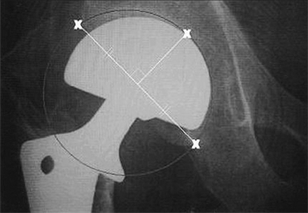
Fig. 1 The image shows how to calculate the acetabular cartilage degeneration. The acetabular bony margin was used instead of the acetabular cup of total hip arthroplasty. Because the shape of the acetabulum is not spherical, three points that could be observed through the AP x-ray findings were selected: the acetabular superior-outer margin, inferior-inner margin and the point where the line that vertically bisects the line that connects the acetabular superior-outer and inferior-inner margin meets the acetabular bony margin. The wear program calculated the circle that passes through these three points.
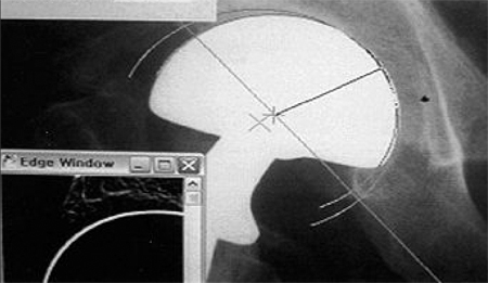
Fig. 2 Image showing the wear analysis procedure, known as the 'Hip 32' (Computer assisted vector wear analysis system, University of Chicago Ortho. Ver. 4.0).7
Reference
-
1. D'Arcy J, Devas M. Treatment of fractures of the femoral neck by replacement with the Thompson prosthesis. J Bone Joint Surg Br. 1976. 58:279–286.2. Harris WH, Rushfeldt PD, Carlson CE, Scholler JM, Mann RW. The hip Society. Pressure distribution in the hip and selection of hemi-arthroplasty. The Hip: Proceedings of the Third Open Scientific Meeting of the Hip Society. 1975. St. Louis: CV Mosby;93–98.3. Hinchey JJ, Day PL. Primary prosthetic replacement in fresh femoral-neck fractures. A review of 294 consecutive cases. J Bone Joint Surg Am. 1964. 46:223–240.4. Jensen JS, Holstein P. A long term follow-up of Moore arthroplasty in femoral neck fractures. Acta Orthop Scand. 1975. 46:764–774.
Article5. Ehrlich MG, Mankin HJ, Jones H, Grossman A, Crispen C, Ancona D. Biochemical confirmation of an experimental osteoarthritis model. J Bone Joint Surg Am. 1975. 57:392–396.
Article6. von Knoch M, Sychterz CJ, Engh CA Jr, Engh CA Sr. Incidence of late bead shedding from uncemented porous coated cups. A radiographic evaluation. Clin Orthop Relat Res. 1997. 342:99–105.7. Ebramzadeh E, Sangiorgio SN, Lattuada F, Kang JS, Chiesa R, Mckellop HA, et al. Accuracy of measurement of polyethylene wear with use of radiographs of total hip replacements. J Bone Joint Surg Am. 2003. 85A:2378–2384.
Article8. Schmalzried TP, Shepherd EF, Dorey FJ, Jackson WO, dela Rosa M, Fa'vae F, et al. The John Charnley Award. Wear is a function of use, not time. Clin Orthop Relat Res. 2000. 381:36–46.9. Cruess RL, Kwok DC, Duc PN, Lecavalier MA, Dang GT. The response of articular cartilage to weight-bearing against metal. A study of hemiarthroplasty of the hip in the dog. J Bone Joint Surg Br. 1984. 66:592–597.
Article10. Minihane KP, Turner TM, Urban RM, Williams JM, Thonar EJ, Sumner DR. Effect of hip hemiarthroplasty on articular cartilage and bone in a canine model. Clin Orthop Relat Res. 2005. 437:157–163.
Article11. van der Meulen MC, Beaupré GS, Smith RL, Giddings VL, Allen WA, Athanasiou KA, et al. Factors influencing changes in articular cartilage following hemiarthroplasty in sheep. J Orthop Res. 2002. 20:669–675.
Article12. Ehrlich MG, Mankin HJ, Jones H, Wright R, Crispen C, Vigliani C. Collagenase and collagenase inhibitors in osteoarthritic and normal cartilage. J Clin Invest. 1977. 59:226–233.
Article13. Kempson GE, Muir H, Pollard C, Tuke M. The tensile properties of the cartilage of human femoral condyles related to the content of collagen and glycosaminoglycans. Biochim Biophys Acta. 1973. 297:456–472.
Article14. Freeman MA. The pathogenesis of primary osteoarthrosis: An hypothesis. Mod Trends Orthop. 1972. 6:40–94.15. McGibbon CA, Krebs DE, Trahan CA, Trippel SB, Mann RW. Cartilage degeneration in relation to repetitive pressure: case study of a unilateral hip hemiarthroplasty patient. J Arthroplasty. 1999. 14:52–58.16. Dalldorf PG, Banas MP, Hicks DG, Pellegrini VD Jr. Rate of degeneration of human acetabular cartilage after hemiarthroplasty. J Bone Joint Surg Am. 1995. 77:877–882.
Article17. Kurrat HJ, Oberländer W. The thickness of the cartilage in the hip joint. J Anat. 1978. 126:145–155.18. Athanasiou KA, Agarwal A, Dzida FJ. Comparative study of the intrinsic mechanical properties of the human acetabular and femoral head cartilage. J Orthop Res. 1994. 12:340–349.
Article19. Eckstein F, von Eisenhart-Rothe R, Landgraf J, Adam C, Loehe F, Müller-Gerbl M, et al. Quantitative analysis of incongruity, contact areas and cartilage thickness in the human hip joint. Acta Anat (Basel). 1997. 158:192–204.
Article
- Full Text Links
-
- Actions
-
Cited
- CITED
-
- Close
- Share
-
- Similar articles
-
- The Reaction of the Acetabular Articular Cartilage to Bipolar Hemiarthroplasty
- Degeneration of the Acetabular Articular Cartilage to Bipolar Hemiarthroplasty
- Analysis of Risk Factors Affecting Degeneration of the Acetabular Cartilage after Bipolar Hemiarthroplasty
- Bipolar Endoprosthesis between Femoral Neck Fracture and Avascular Necrosis of Femoral Head: Comparison of the Acetabular Migration and Function
- Histological and Histochemical Study of the Acetabular Articular Cartilage in Avascular Necrosis of Femoral Head
