Three Cases of Diffuse Large B-Cell Lymphoma Presenting as Primary Splenic Lymphoma
- Affiliations
-
- 1Department of Internal Medicine, Yonsei University College of Medicine, Seoul, Korea. medi@yumc.yonsei.ac.kr
- 2Department of Hematology-Oncology, Yonsei University College of Medicine, Seoul, Korea.
- 3Department of Radiation Oncology, Yonsei University College of Medicine, Seoul, Korea.
- 4Department of Pathology, Yonsei University College of Medicine, Seoul, Korea.
- KMID: 2158153
- DOI: http://doi.org/10.3349/ymj.2005.46.5.703
Abstract
- Primary splenic lymphoma (PSL) is often defined as generalized lymphoma with splenic involvement as the dominant feature. It is a rare disease that comprises approximately 1% of all malignant lymphomas. We investigated three cases of non-Hodgkin's splenic lymphoma that had different clinical features on presentation. The patients' survival times from diagnosis ranged from 59 to 143 months, without evidence of relapse after splenectomy and chemotherapy, with or without radiotherapy. This data suggest that PSL is potentially curable. Further studies are needed to evaluate the impact that different treatment modalities without splenectomy have on patient survival.
Keyword
MeSH Terms
Figure
-
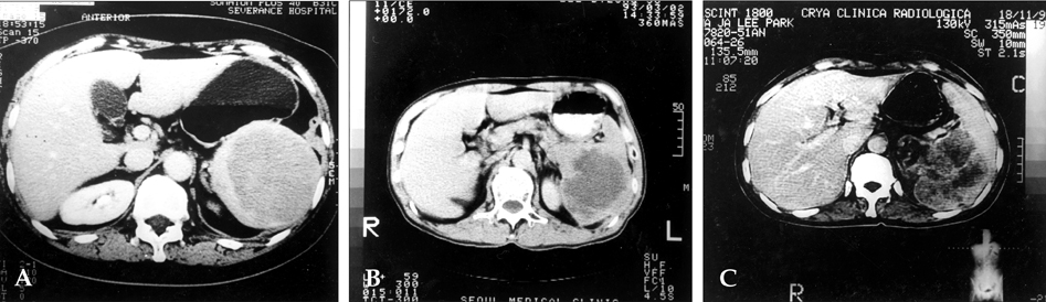
Fig. 1 Computed tomography (CT) scans of each case. A) Case 1- a huge splenic mass with homogenous high-density areas, including an area of low-density without perisplenic spread. B) Case 2- a well-localized and capsulated splenic mass with internal necrosis or hemorrhage and partial bulging of the pancreatic tail portion at the junctional lesion with spleen. C) Case 3- a heterogeneous, lobulated splenic mass measuring 10×8×8 cm with bulging contours, necrotic foci and focal calcification.
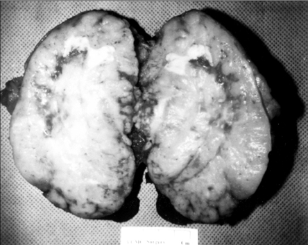
Fig. 2 Gross examination of the resected spleen of Case 1. The exposed cut surface shows a bulging, well-demarcated grayish-yellow nodular solid mass, measuring 12 cm in the largest dimension. Multifocal grayish-yellow necrotic foci are present. The splenic parenchyma is nearly replaced by this lesion.
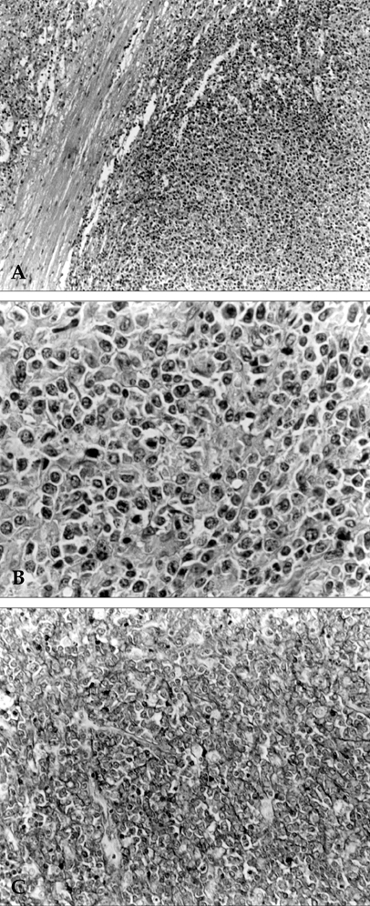
Fig. 3 Histopathologic findings of Case 1. A) Mass border on low power (×100) showing an expanding growth pattern of tumor cells. B) Mass on high power (×400) showing non-Hodgkin's lymphoma, diffuse large B-cell type. Tumor cells have large nuclei, open chromatin, and prominent nucleoli. C) Immunohistochemical stain for L26 (B-cell marker) (×200) showing prominent immunopositivity to the tumor cells.
Reference
-
1. Ahmann DL, Kiely JM, Harrison EG, Payne WS. Malignant lymphoma of the spleen. Cancer. 1966. 19:461–469.2. Kim KH, Cho CK, Choo SW, Kim HJ, Kim KS. Primary lymphoma of the spleen- a case report. Korean J Surg. 1997. 52:912–917.3. Hahn JS, Lee S, Chong SY, Min YH, Ko YW. Eight-year experience of malignant lymphoma-survival and prognostic factors. Yonsei Med J. 1997. 38:270–284.4. Fawcett DW. A textbook of histology. 1994. 12th ed. New York: Chapman & Hall;460–472.5. Cotran RS, Kumar V, Robbins SL. Pathologic basis of disease. 1994. 5th ed. Philadelphia: W.B. Saunders;667–671.6. McCormick WF, Kashgarian M. The weight of the adult human spleen. Am J Clin Pathol. 1965. 43:332–333.7. Gobbi PG, Grignani GE, Pozzetti U, Bertoloni D, Pieresca C, Montagna G, et al. Primary splenic lymphoma: Does it exist? Haematologica. 1994. 79:286–293.8. Kraemer BB, Osborne BM, Butler JJ. Primary splenic presentation of malignant lymphoma and related disorders-a study of 49 cases. Cancer. 1984. 54:1606–1619.9. Kehoe J, Straus DJ. Primary lymphoma of the spleen-clinical features and outcome after splenectomy. Cancer. 1988. 62:1433–1438.10. Falk S, Stutte HJ. Primary malignant lymphomas of the spleen. Cancer. 1990. 66:2612–2619.11. Dasgupta T, Coombes BC, Brasfield RD. Primary malignant neoplasms of the spleen. Surg Gynecol Obstet. 1965. 120:947–960.12. Skarin AT, Davey FR, Moloney WC. Lymphosarcoma of the spleen. Arch Intern Med. 1971. 127:259–265.13. Dachman AH, Buck JL, Krishnan J, Aguilera NS, Buetow PC. Primary non-Hodgkin's splenic lymphoma. Clin Radiol. 1998. 53:137–142.14. Xiros N, Economopoulos T, Christodoulidis C, Dervenoulas J, Papageorgiou E, Mellou S, et al. Splenectomy in patients with malignant non-Hodgkin's lymphoma. Eur J Haematol. 2000. 64:145–150.15. Brox A, Bishinsky JI, Berry G. Primary non-Hodgkin lymphoma of the spleen. Am J Hematol. 1991. 38:95–100.16. Karpeh MS Jr, Hicks DG, Torosian MH. Colon invasion by primary splenic lymphoma: a case report and review of the literature. Surgery. 1992. 111:224–227.17. Morel P, Dupriez B, Gosselin B, Fenaux P, Estienne MH, Facon T, et al. Role of early splenectomy in malignant lymphomas with prominent splenic involvement (Primary lymphomas of the spleen)- A study of 59 cases. Cancer. 1993. 71:207–215.18. Lee JD, Park CH, Griffith J, Vernick J. Gallium-67 scintiscan in the diagnosis of primary splenic non-Hodgkin's lymphoma after the treatment of Hodgkin's disease. J Nucl Med. 1992. 33:1183–1185.19. Harris NL, Aisengerg AC, Meyer JE, Ellman L, Elman A. Diffuse large cell (histiocytic) lymphoma of the spleen. Cancer. 1984. 54:2460–2467.20. Abraksia S, Dileep KP, Kasal J. Two unusual lymphomas. J Clin Oncol. 2000. 18:3731–3733.
- Full Text Links
-
- Actions
-
Cited
- CITED
-
- Close
- Share
-
- Similar articles
-
- Primary Splenic Lymphoma with Splenic Hilar Lymphadenopathy
- Relapse of Ocular Lymphoma following Primary Testicular Diffuse Large B-cell Lymphoma
- Diffuse Large B-Cell Lymphoma in the Portal Vein
- Primary Cutaneous T-cell/histiocyte-rich B-cell Lymphoma
- Two Cases of Primary Esophageal Diffuse Large B Cell Lymphoma: Therapeutic Considerations and a Literature Review
