Ann Dermatol.
2011 Sep;23(Suppl 1):S116-S118. 10.5021/ad.2011.23.S1.S116.
A Case of a Subepidermal Calcified Nodule on the Sole without Trauma
- Affiliations
-
- 1Department of Dermatology, College of Medicine, Hallym University, Seoul, Korea. dermap@paran.com
- 2Department of Pathology, College of Medicine, Hallym University, Seoul, Korea.
- KMID: 2156767
- DOI: http://doi.org/10.5021/ad.2011.23.S1.S116
Abstract
- Subepidermal calcified nodule is an uncommon form of calcinosis cutis, which most commonly occurs in children. It usually presents as an asymptomatic, solitary verrucous nodule on the head and neck region, but occasionally as multiple lesions. Serum calcium and phosphorus levels are usually normal. Histopathology shows well-formed homogeneous eosinophilic material and granules in the upper dermis. Material in the dermis stained with von Kossa was positive. We report on an unusual case of a subepidermal calcified nodule occurring on the sole. A 21-month-old male presented with an oval-shaped, whitish, hard nodule measuring 5x5 mm on the left sole, without any previous history of trauma.
Keyword
MeSH Terms
Figure
-
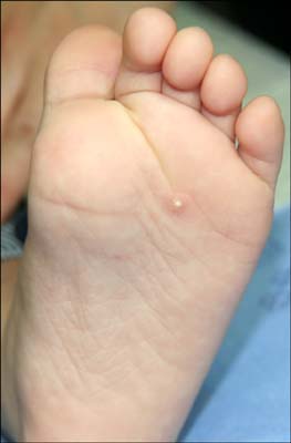
Fig. 1 An oval-shaped, whitish papule measuring 0.5×0.5 cm on an erythematous base of the left sole.
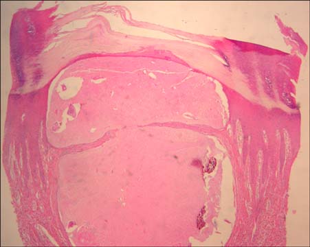
Fig. 2 Haematoxylin and eosin-stained sections showed a hyperkeratotic and acanthotic epidermis overlying a cystic structure in the upper dermis. The cystic space contained amorphous eosinophilic materials as multilobulated masses (H&E stain, ×40).

Fig. 3 Amorphous black to dark brown colored materials were confirmed as calcium by staining with von Kossa (von Kossa stain, ×40).
Reference
-
1. Elder DE, Elenitsas R, Johnson BL Jr, Murphy GF, Xu G. Lever's histopathology of the skin. 2009. 10th ed. Philadelphia: Lippincott Williams & Wilkins;435–438.2. Baselga E, Fairley JA. Harper J, Oranje A, Prose N, editors. Calcification and ossification in the skin. Textbook of pediatric dermatology. 2000. Oxford: Blackwell Science;788–789. .
Article3. Rho NK, Youn SJ, Park HS, Kim WS, Lee ES. Calcified nodule on the heel of a child following a single heel stick in the neonatal period. Clin Exp Dermatol. 2003. 28:502–503.
Article4. Hyun JS, Park CJ, Yi JY. A case of calcinosis cutis following heel sticks. Korean J Dermatol. 2000. 38:1270–1272.5. Cambiaghi S, Restano L, Imondi D. Calcified nodule of the heel. Pediatr Dermatol. 1997. 14:494.6. Williamson D, Holt PJ. Calcified cutaneous nodules on the heels of children: a complication of heel sticks as a neonate. Pediatr Dermatol. 2001. 18:138–140.
Article7. Lemont H, Brady J. Infant heel nodules. Calcification of epidermal cysts. J Am Podiatr Med Assoc. 2002. 92:112–113.8. Son SJ, Kim WS. A case of subepidermal calcified nodules. Korean J Dermatol. 1976. 14:173–178.9. Paek SH, Kim YH, Kim DW, Jun JB, Chung SL. Subepidermal calcified nodule. Ann Dermatol. 1996. 8:271–296.
Article10. Jung GD, Choi YH, Jeon YM, Song ES. Subepidermal calcified nodule arising in the lesion of clear cell syringoma. Korean J Dermatol. 2000. 38:1660–1663.11. Kim YK, Choi YH, Choi KC, Kim HK. A case of subepidermal calcified nodules showing an unusual clinical manifestation. Korean J Dermatol. 1983. 21:595–599.12. Neidner KH. Light microscopic findings. Clinics in Dermatology. 1988. 6:93–99.
Article13. Barson AJ, Symonds J. Calcified pituitary concretions in the newborn. Arch Dis Child. 1997. 52:642–645.
Article14. Winer LH. Solitary congenital nodular calcification of the skin. AMA Arch Derm Syphilol. 1952. 66:204–211.
Article15. Woods B, Kellaway TD. Cutaneous calculi subepidermal calcified nodules. Br J Dermatol. 1963. 75:1–11.16. Plott T, Wiss K, Raimer SS, Solomon AR. Recurrent subepidermal calcified nodule of nose. Pediatr Dermatol. 1988. 5:107–111.17. Shmunes E, Wood MG. Subepidermal calcified nodules. Arch Dermatol. 1972. 105:593–597.
Article18. Evans MJ, Blessing K, Gray ES. Subepidermal calcified nodule in children: a clinicopathologic study of 21 cases. Pediatr Dermatol. 1995. 12:307–310.
Article19. Tezuka T. Cutaneous calculus - its pathogenesis. Dermatologica. 1980. 161:191–199.
Article20. Evans LA, Evans CM, Cobb MW. An asymptomatic papule on the face. Pediatr Dermatol. 1996. 13:253–254.
Article21. Lee SS, Felsenstein J, Tanzer FR. Calcinosis cutis circumscripta. Treatment with an intralesional corticosteroid. Arch Dermatol. 1978. 114:1080–1081.
Article22. Jun JH, Lee JB, Kim SJ, Lee SC, Won YH. A case of subepidermal calcified nodule with transepidermal elimination. Korean J Dermatol. 2003. 41:89–91.23. Kim DH, Ham SH, Kang H, Cho SH, Park YM. Subepidermal calcified nodule of the buttock. Ann Dermatol. 2000. 12:74–76.
Article24. Lee WC, Cha YC, Lee SJ, Na GY, Kim DW, Lee SK. Subepidermal calcified nodule of the finger. Korean J Dermatol. 2003. 41:1414–1416.
