A Case of Dedifferentiated LiposarcomaThat Developed in the Dermis
- Affiliations
-
- 1Department of Dermatology, School of Medicine, Ewha Womans University, Seoul, Korea. kbmyung@ewha.ac.kr
- KMID: 2156395
- DOI: http://doi.org/10.5021/ad.2008.20.4.204
Abstract
- Dedifferentiated liposarcoma is a variant of liposarcoma, and this is characterized by the coexistence of well-differentiated liposarcoma with areas of poorly differentiated, non-lipogenic tumor and this is also known to be associated with more aggressive behavior. Dedifferentiated liposarcoma occurs principally in the retroperitoneum or the deep soft tissue of limbs, but it can also occur in subcutaneous locations. We report here on a peculiar case of dedifferentiated liposarcoma that developed in the dermis, which is an exceedingly rare location for this type of tumor. The occurrence of this tumor in the dermis made it easy to surgically remove and monitor for recurrence, and we expect this patient to have a better prognosis than that of a patient with dedifferentiated liposarcoma located in the retroperitoneum or deep soft tissue.
Keyword
MeSH Terms
Figure
-
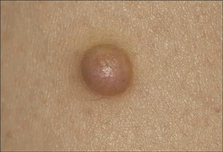
Fig. 1 A 9 mm-size dome-shape brownish nodule was located on the right thigh.
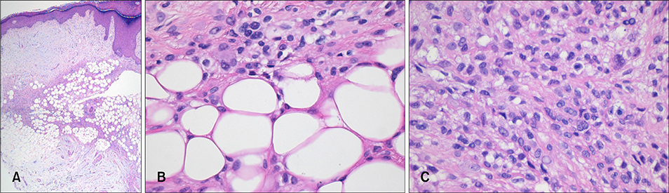
Fig. 2 (A) Well-differentiated elements consisting of mature adipocytes and lipoblasts were comingled with dedifferentiated elements composed of poorly differentiated round cells in the myxoid dermis (H&E, ×40). (B) Mature adipocytes and well-differentiated lipoblasts were observed in the well-differentiated areas. Some of these cells showed enlarged hyperchromatic atypical nuclei (H&E, ×400). (C) The dedifferentiated areas consisted of poorly differentiated, slightly lipogenic or nonlipogenic round cells (H&E, ×400).

Fig. 3 (A) A nonlipogenic lipoblast exhibited a small number of non-membrane bound lipid inclusions (EM, ×12,000). (B) A slightly lipogenic lipoblast showed multiple lipid inclusions (EM, ×7,000). (C) The cell with a large lipid vacuole corresponded to a signet ring cell noted on the light microscopic examination (EM, ×2,000).
Cited by 1 articles
-
Subcutaneous Myxoid and Round Cell Liposarcoma
Hyun Soo Roh, Ha Eun Lee, Moon Hyang Park, Joo Yeon Ko, Young Suck Ro
Ann Dermatol. 2011;23(3):338-341. doi: 10.5021/ad.2011.23.3.338.
Reference
-
1. Fletcher CDM. Adipocytic tumors. In : Fletcher CDM, editor. Diagnostic histopathology of tumors. 2nd ed. London: Churchill Livingstone;2000. p. 1474–1485.2. Dei Tos AP, Mentzel T, Fletcher CD. Primary liposarcoma of the skin: a rare neoplasm with unusual high grade features. Am J Dermatopathol. 1998; 20:332–338.
Article3. Weiss SW. WHO histological typing of soft tissue tumours. 2nd ed. Berlin: Springer-Verlag;1994.4. Kim TH, Kim JW, Kim SW, Kim DS, Ahn KY. Myxoid liposarcoma. Ann Dermatol. 1996; 8:141–148.
Article5. Kim WH, Seung NR, Kim CW, Cho HJ, Kim KH, Kim KJ. A case of well-differentiated liposarcoma. Ann Dermatol. 2006; 18:105–108.
Article6. Dei Tos AP. Liposarcoma: new entities and evolving concepts. Ann Diagn Pathol. 2000; 4:252–266.
Article7. Fletcher CD, Akerman M, Dal Cin P, de Wever I, Mandahl N, Mertens F, et al. Correlation between clinicopathological features and karyotype in lipomatous tumors. A report of 178 cases from the Chromosomes and Morphology (CHAMP) Collaborative Study Group. Am J Pathol. 1996; 148:623–630.8. Turc-Carel C, Limon J, Dal Cin P, Rao U, Karakousis C, Sandberg AA. Cytogenetic studies of adipose tissue tumors. II. Recurrent reciprocal translocation t(12;16)(q13;p11) in myxoid liposarcomas. Cancer Genet Cytogenet. 1986; 23:291–299.
Article9. Crozat A, Aman P, Mandahl N, Ron D. Fusion of CHOP to a novel RNA-binding protein in human myxoid liposarcoma. Nature. 1993; 363:640–644.
Article10. Rabbitts TH, Forster A, Larson R, Nathan P. Fusion of the dominant negative transcription regulator CHOP with a novel gene FUS by translocation t(12;16) in malignant liposarcoma. Nat Genet. 1993; 4:175–180.
Article11. Knight JC, Renwick PJ, Dal Cin P, Van den Berghe H, Fletcher CD. Translocation t(12;16)(q13;p11) in myxoid liposarcoma and round cell liposarcoma: molecular and cytogenetic analysis. Cancer Res. 1995; 55:24–27.12. Dal Cin P, Sciot R, Panagopoulos I, Aman P, Samson I, Mandahl N, et al. Additional evidence of a variant translocation t(12;22) with EWS/CHOP fusion in myxoid liposarcoma: clinicopathological features. J Pathol. 1997; 182:437–441.
Article13. Enzinger FM, Weiss SW. Liposarcomas. In : Enzinger FM, Weiss SW, editors. Soft tissue tumors. 3rd ed. St. Louis: Mosby;1995. p. 431–466.14. Meis JM. "Dedifferentiation" in bone and soft-tissue tumors. A histological indicator of tumor progression. Pathol Annu. 1991; 26:37–62.15. Henricks WH, Chu YC, Goldblum JR, Weiss SW. Dedifferentiated liposarcoma: a clinicopathological analysis of 155 cases with a proposal for an expanded definition of dedifferentiation. Am J Surg Pathol. 1997; 21:271–281.16. Evans HL, Khurana KK, Kemp BL, Ayala AG. Heterologous elements in the dedifferentiated component of dedifferentiated liposarcoma. Am J Surg Pathol. 1994; 18:1150–1157.
Article17. Brooks JJ, Connor AM. Atypical lipoma of the extremities and peripheral soft tissues with dedifferentiation: implications for management. Surg Pathol. 1990; 3:169–178.18. Evans HL. Liposarcomas and atypical lipomatous tumors: a study of 66 cases followed for a minimum of 10 years. Surg Pathol. 1988; 1:41–54.19. Lucas DR, Nascimento AG, Sanjay BK, Rock MG. Well-differentiated liposarcoma. The Mayo Clinic experience with 58 cases. Am J Clin Pathol. 1994; 102:677–683.
Article20. McCormick D, Mentzel T, Beham A, Fletcher CD. Dedifferentiated liposarcoma. Clinicopathologic analysis of 32 cases suggesting a better prognostic subgroup among pleomorphic sarcomas. Am J Surg Pathol. 1994; 18:1213–1223.
Article
- Full Text Links
-
- Actions
-
Cited
- CITED
-
- Close
- Share
-
- Similar articles
-
- Dedifferentiated Chordoma: Report of a case
- Dedifferentiated Liposarcoma with a Peculiar Whorling Pattern: A Case Report
- Dedifferentiated Liposarcoma in the Thigh: Case Report
- A Dedifferentiated Liposarcoma of Soft Tissue with Features of Fibrosarcomatous Redifferentiation
- Giant Liposarcoma Arising in the Mesentery
