Physiological and Functional Magnetic Resonance Imaging Using Balanced Steady-state Free Precession
- Affiliations
-
- 1Magnetic Resonance Imaging Lab, Department of Bio and Brain Engineering, Korea Advanced Institute of Science and Technology, Daejeon 305-701, Korea. sunghongpark@kaist.ac.kr
- 2Department of Radiology, Seoul National University College of Medicine, Seoul 110-744, Korea.
- KMID: 2155524
- DOI: http://doi.org/10.3348/kjr.2015.16.3.550
Abstract
- Balanced steady-state free precession (bSSFP) is a highly efficient pulse sequence that is known to provide the highest signal-to-noise ratio per unit time. Recently, bSSFP is getting increasingly popular in both the research and clinical communities. This review will be focusing on the application of the bSSFP technique in the context of probing the physiological and functional information. In the first part of this review, the basic principles of bSSFP are briefly covered. Afterwards, recent developments related to the application of bSSFP, in terms of physiological and functional imaging, are introduced and reviewed. Despite its long development history, bSSFP is still a promising technique that has many potential benefits for obtaining high-resolution physiological and functional images.
Keyword
MeSH Terms
Figure
-
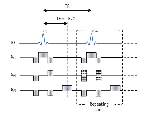
Fig. 1 Pulse sequence diagram of balanced steady-state free precession. Sum of all gradients in each of three directions (slice-selection, phase-encoding, and frequency-encoding) is zero. Phase of radio frequency (RF) pulse, denoted as phase-cycling angle (θ), can be incremented after every repetition time (TR). Echo time (TE) is typically set to half of TR. GSS, GPE, and GFE represent gradients for slice selection, phase encoding, and frequency encoding, respectively.

Fig. 2 Magnetization changes of balanced steady-state free precession (bSSFP) sequence with half-alpha preparation and phase-cycling angle of 180°. Mz denotes longitudinal magnetization and My denotes transverse magnetization along y-axis in rotating frame, and α denotes flip angle of radio frequency pulse. Notice that magnitude of transvers

Fig. 3 Balanced steady-state signal profile as function of off-resonance frequency in one repetition time. Magnitude and phase responses of balanced steady-state free precession (bSSFP) signals are shown as function of off-resonance precession angle per each repetition time. bSSFP signal profile for phase-cycling angles of 0° (A), 90° (B), 180° (C), and 270° (D) are shown. Notice that signal profile is periodic and that signal profile shifts with each phase-cycling angle.
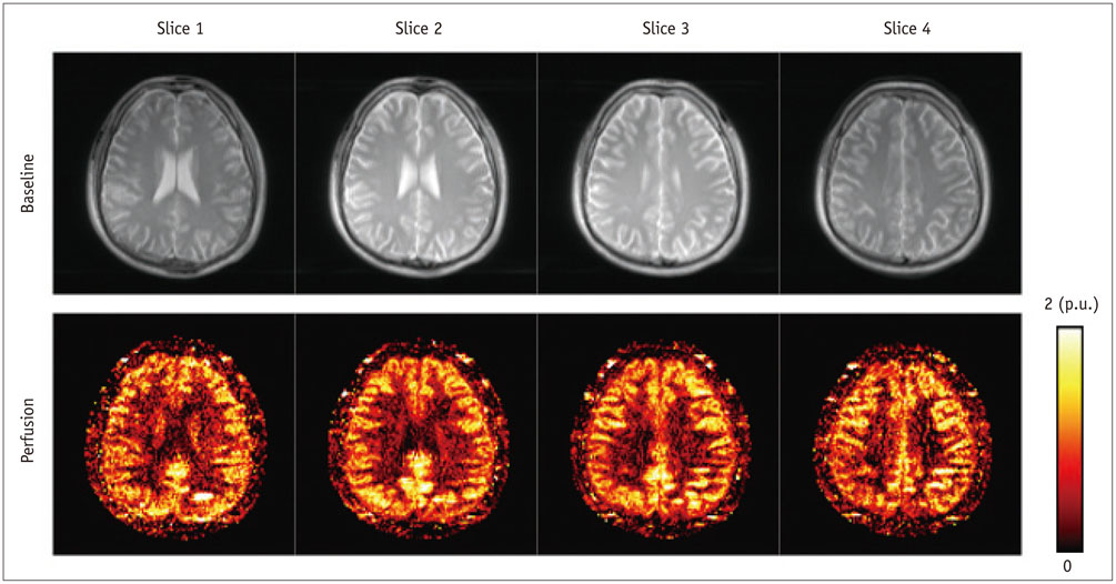
Fig. 4 Baseline and cerebral blood flow images of brain and were acquired using three-dimensional pseudo-continuous arterial spin labeling technique with balanced steady-state free precession readout scheme. Repetition time/echo time = 4/2 ms, field of view = 240 × 180 × 20 mm3, matrix = 128 × 96 × 4, number of average for pair-wise subtraction = 40, and scan time = 4 minutes. p.u. = percent unit

Fig. 5 Inter-slice blood flow images acquired with alternate ascending/descending directional navigation (ALADDIN). Balanced steady-state free precession sequence is used for ALADDIN acquisition. Baseline (A), perfusion with direction from feet (F) to head (H) (B), and perfusion with direction from head (H) to feet (F) (C) images are shown. Arrow indicates superior sagittal sinus. Repetition time/echo time = 4/2 ms, field of view = 230 × 230 mm2, matrix = 128 × 128, slice thickness = 5 mm, phase oversampling = 50%, number of slices = 15, number average for pair-wise subtraction = 8, and scan time = -3 minutes. p.u. = percent unit
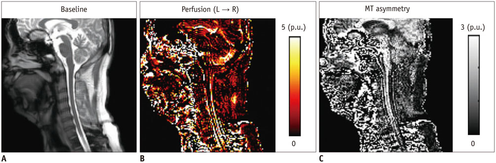
Fig. 6 Sagittal view of inter-slice blood flow and magnetization transfer (MT) asymmetry images in cervical spines simultaneously acquired with alternate ascending/descending directional navigation (ALADDIN). Balanced steady-state free precession sequence is used for ALADDIN acquisition. Baseline (A), perfusion (B), and MT asymmetry (C) images are shown. Repetition time/echo time = 4/2 ms, field of view = 240 × 240 mm2, matrix = 128 × 128, slice thickness = 4 mm, phase oversampling = 50%, number of slices = 13, number average for pair-wise subtraction = 8, and scan time = -3 minutes. p.u. = percent unit
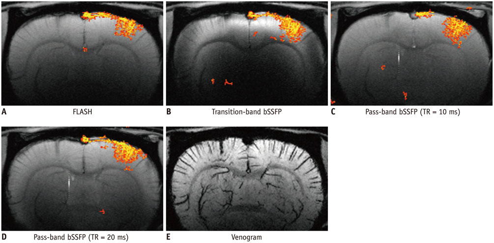
Fig. 7 Investigation of activation foci of high-resolution functional MRI maps. Functional MRI maps acquired at 9.4-T with fast low angle shot (FLASH) (A), transition-band balanced steady-state free precession (bSSFP) with time to repeat (TR) = 20 ms (B), pass-band bSSFP with TR = 10 ms (C), and pass-band bSSFP with TR = 20 ms (D). Echo time was set to half of TR for all images. Each functional MRI map is overlaid on corresponding averaged baseline image. Matrix size = 256 × 128, field of view = 2.2 × 1.1 cm2, slice thickness = 1 mm, and number of slice = 1 for all images. Flip angle was 8°, 4°, 16°, and 16° for images in A-D, respectively, and angles were optimized based on simulation at 9.4-T. E. Venogram with minimum intensity projection at same location as functional MRI maps in A-D.
Cited by 3 articles
-
Left Gastric Vein Visualization with Hepatopetal Flow Information in Healthy Subjects Using Non-Contrast-Enhanced Magnetic Resonance Angiography with Balanced Steady-State Free-Precession Sequence and Time-Spatial Labeling Inversion Pulse
Akihiro Furuta, Hiroyoshi Isoda, Tsuyoshi Ohno, Ayako Ono, Rikiya Yamashita, Shigeki Arizono, Aki Kido, Naotaka Sakashita, Kaori Togashi
Korean J Radiol. 2018;19(1):32-39. doi: 10.3348/kjr.2018.19.1.32.Brain Regional Homogeneity Changes in Cirrhotic Patients with or without Hepatic Encephalopathy Revealed by Multi-Frequency Bands Analysis Based on Resting-State Functional MRI
Gaoyan Zhang, Yue Cheng, Wen Shen, Baolin Liu, Lixiang Huang, Shuangshuang Xie
Korean J Radiol. 2018;19(3):452-462. doi: 10.3348/kjr.2018.19.3.452.Evaluation of Tumor Blood Flow Using Alternate Ascending/Descending Directional Navigation in Primary Brain Tumors: A Comparison Study with Dynamic Susceptibility Contrast Magnetic Resonance Imaging
Hyeree Park, Joonhyuk Lee, Sung-Hong Park, Seung Hong Choi
Korean J Radiol. 2019;20(2):275-282. doi: 10.3348/kjr.2018.0300.
Reference
-
1. Alsop DC. Phase insensitive preparation of single-shot RARE: application to diffusion imaging in humans. Magn Reson Med. 1997; 38:527–533.2. Feinberg DA, Kiefer B, Johnson G. GRASE improves spatial resolution in single shot imaging. Magn Reson Med. 1995; 33:529–533.3. Crelier GR, Hoge RD, Munger P, Pike GB. Perfusion-based functional magnetic resonance imaging with single-shot RARE and GRASE acquisitions. Magn Reson Med. 1999; 41:132–136.4. Carr HY. Steady-state free precession in nuclear magnetic resonance. Phys Rev. 1958; 112:1693–1701.5. Zur Y, Stokar S, Bendel P. An analysis of fast imaging sequences with steady-state transverse magnetization refocusing. Magn Reson Med. 1988; 6:175–193.6. Oppelt A, Graumann R, Barfuss H, Fischer H, Hartl W, Shajor W. FISP-a new fast MRI sequence. Electromedica. 1986; 54:15–18.7. Scheffler K, Lehnhardt S. Principles and applications of balanced SSFP techniques. Eur Radiol. 2003; 13:2409–2418.8. Miller KL. FMRI using balanced steady-state free precession (SSFP). Neuroimage. 2012; 62:713–719.9. Bieri O, Scheffler K. Fundamentals of balanced steady state free precession MRI. J Magn Reson Imaging. 2013; 38:2–11.10. Miller KL, Tijssen RHN, Stikov N, Okell TW. Steady-state MRI: methods for neuroimaging. Imaging Med. 2011; 3:93–105.11. Scheffler K, Hennig J. Is TrueFISP a gradient-echo or a spin-echo sequence? Magn Reson Med. 2003; 49:395–397.12. Carr JC, Simonetti O, Bundy J, Li D, Pereles S, Finn JP. Cine MR angiography of the heart with segmented true fast imaging with steady-state precession. Radiology. 2001; 219:828–834.13. Hays AG, Schär M, Kelle S. Clinical applications for cardiovascular magnetic resonance imaging at 3 tesla. Curr Cardiol Rev. 2009; 5:237–242.14. Kim SG. Quantification of relative cerebral blood flow change by flow-sensitive alternating inversion recovery (FAIR) technique: application to functional mapping. Magn Reson Med. 1995; 34:293–301.15. Martirosian P, Klose U, Mader I, Schick F. FAIR true-FISP perfusion imaging of the kidneys. Magn Reson Med. 2004; 51:353–361.16. Boss A, Martirosian P, Graf H, Claussen CD, Schlemmer HP, Schick F. High resolution MR perfusion imaging of the kidneys at 3 Tesla without administration of contrast media. Rofo. 2005; 177:1625–1630.17. Fenchel M, Martirosian P, Langanke J, Giersch J, Miller S, Stauder NI, et al. Perfusion MR imaging with FAIR true FISP spin labeling in patients with and without renal artery stenosis: initial experience. Radiology. 2006; 238:1013–1021.18. Boss A, Martirosian P, Klose U, Nägele T, Claussen CD, Schick F. FAIR-TrueFISP imaging of cerebral perfusion in areas of high magnetic susceptibility differences at 1.5 and 3 Tesla. J Magn Reson Imaging. 2007; 25:924–931.19. Ludescher B, Martirosian P, Klose U, Nägele T, Schick F, Ernemann U. Determination of the rCBF in the amygdala and rhinal cortex using a FAIR-TrueFISP sequence. Korean J Radiol. 2011; 12:554–558.20. Zun Z, Wong EC, Nayak KS. Assessment of myocardial blood flow (MBF) in humans using arterial spin labeling (ASL): feasibility and noise analysis. Magn Reson Med. 2009; 62:975–983.21. Boss A, Martirosian P, Claussen CD, Schick F. Quantitative ASL muscle perfusion imaging using a FAIR-TrueFISP technique at 3.0 T. NMR Biomed. 2006; 19:125–132.22. Buchbender S, Obenauer S, Mohrmann S, Martirosian P, Buchbender C, Miese FR, et al. Arterial spin labelling perfusion MRI of breast cancer using FAIR TrueFISP: initial results. Clin Radiol. 2013; 68:e123–e127.23. Dai W, Garcia D, de Bazelaire C, Alsop DC. Continuous flowdriven inversion for arterial spin labeling using pulsed radio frequency and gradient fields. Magn Reson Med. 2008; 60:1488–1497.24. Wu WC, Fernández-Seara M, Detre JA, Wehrli FW, Wang J. A theoretical and experimental investigation of the tagging efficiency of pseudocontinuous arterial spin labeling. Magn Reson Med. 2007; 58:1020–1027.25. Park SH, Wang DJ, Duong TQ. Balanced steady state free precession for arterial spin labeling MRI: initial experience for blood flow mapping in human brain, retina, and kidney. Magn Reson Imaging. 2013; 31:1044–1050.26. Wu WC, Jain V, Li C, Giannetta M, Hurt H, Wehrli FW, et al. In vivo venous blood T1 measurement using inversion recovery true-FISP in children and adults. Magn Reson Med. 2010; 64:1140–1147.27. Hori M, Shiraga N, Watanabe Y, Aoki S, Isono S, Yui M, et al. Time-resolved three-dimensional magnetic resonance digital subtraction angiography without contrast material in the brain: initial investigation. J Magn Reson Imaging. 2009; 30:214–218.28. Yan L, Li C, Kilroy E, Wehrli FW, Wang DJ. Quantification of arterial cerebral blood volume using multiphase-balanced SSFP-based ASL. Magn Reson Med. 2012; 68:130–139.29. Yan L, Wang S, Zhuo Y, Wolf RL, Stiefel MF, An J, et al. Unenhanced dynamic MR angiography: high spatial and temporal resolution by using true FISP-based spin tagging with alternating radiofrequency. Radiology. 2010; 256:270–279.30. Park SH, Duong TQ. Alternate ascending/descending directional navigation approach for imaging magnetization transfer asymmetry. Magn Reson Med. 2011; 65:1702–1710.31. Park SH, Duong TQ. Brain MR perfusion-weighted imaging with alternate ascending/descending directional navigation. Magn Reson Med. 2011; 65:1578–1591.32. Park SH, Zhao T, Kim JH, Boada FE, Bae KT. Suppression of effects of gradient imperfections on imaging with alternate ascending/descending directional navigation. Magn Reson Med. 2012; 68:1600–1606.33. Hodnett PA, Koktzoglou I, Davarpanah AH, Scanlon TG, Collins JD, Sheehan JJ, et al. Evaluation of peripheral arterial disease with nonenhanced quiescent-interval single-shot MR angiography. Radiology. 2011; 260:282–293.34. Edelman RR, Sheehan JJ, Dunkle E, Schindler N, Carr J, Koktzoglou I. Quiescent-interval single-shot unenhanced magnetic resonance angiography of peripheral vascular disease: technical considerations and clinical feasibility. Magn Reson Med. 2010; 63:951–958.35. Santini F, Wetzel SG, Bock J, Markl M, Scheffler K. Time-resolved three-dimensional (3D) phase-contrast (PC) balanced steady-state free precession (bSSFP). Magn Reson Med. 2009; 62:966–974.36. Scheffler K, Seifritz E, Bilecen D, Venkatesan R, Hennig J, Deimling M, et al. Detection of BOLD changes by means of a frequency-sensitive trueFISP technique: preliminary results. NMR Biomed. 2001; 14:490–496.37. Miller KL, Hargreaves BA, Lee J, Ress D, deCharms RC, Pauly JM. Functional brain imaging using a blood oxygenation sensitive steady state. Magn Reson Med. 2003; 50:675–683.38. Miller KL, Smith SM, Jezzard P, Pauly JM. High-resolution FMRI at 1.5T using balanced SSFP. Magn Reson Med. 2006; 55:161–170.39. Lee J, Santos JM, Conolly SM, Miller KL, Hargreaves BA, Pauly JM. Respiration-induced B0 field fluctuation compensation in balanced SSFP: real-time approach for transition-band SSFP fMRI. Magn Reson Med. 2006; 55:1197–1201.40. Lee J, Shahram M, Schwartzman A, Pauly JM. Complex data analysis in high-resolution SSFP fMRI. Magn Reson Med. 2007; 57:905–917.41. Wu ML, Wu PH, Huang TY, Shih YY, Chou MC, Liu HS, et al. Frequency stabilization using infinite impulse response filtering for SSFP fMRI at 3T. Magn Reson Med. 2007; 57:369–379.42. Bowen CV, Menon RS, Gati JS. High field balanced-SSFP fMRI: a BOLD technique with excellent tissue sensitivity and superior large vessel suppression. Proc Intl Soc Mag Reson Med. 2005; 119.43. Lee JH, Dumoulin SO, Saritas EU, Glover GH, Wandell BA, Nishimura DG, et al. Full-brain coverage and high-resolution imaging capabilities of passband b-SSFP fMRI at 3T. Magn Reson Med. 2008; 59:1099–1110.44. Miller KL, Smith SM, Jezzard P, Wiggins GC, Wiggins CJ. Signal and noise characteristics of SSFP FMRI: a comparison with GRE at multiple field strengths. Neuroimage. 2007; 37:1227–1236.45. Zhong K, Leupold J, Hennig J, Speck O. Systematic investigation of balanced steady-state free precession for functional MRI in the human visual cortex at 3 Tesla. Magn Reson Med. 2007; 57:67–73.46. Bowen C, Mason J, Menon R, Gati J. High field balanced-SSFP fMRI: examining a diffusion contrast mechanism using varied flip-angles. Proc 14th ISMRM. Seattle: 2006. p. 665.47. Miller KL, Jezzard P. Modeling SSFP functional MRI contrast in the brain. Magn Reson Med. 2008; 60:661–673.48. Kim TS, Lee J, Lee JH, Glover GH, Pauly JM. Analysis of the BOLD Characteristics in Pass-Band bSSFP fMRI. Int J Imaging Syst Technol. 2012; 22:23–32.49. Patterson S, Beyea S, Bowen C. Quantification of the BOLD contrast mechanism, including its dynamic approach to steady state, for pass-band balanced-SSFP fMRI. Proc Intl Soc Mag Reson Med. Toronto: 2008. p. 2382.50. Park SH, Kim T, Wang P, Kim SG. Sensitivity and specificity of high-resolution balanced steady-state free precession fMRI at high field of 9.4T. Neuroimage. 2011; 58:168–176.51. Park SH, Masamoto K, Hendrich K, Kanno I, Kim SG. Imaging brain vasculature with BOLD microscopy: MR detection limits determined by in vivo two-photon microscopy. Magn Reson Med. 2008; 59:855–865.52. Cheng JS, Gao PP, Zhou IY, Chan RW, Chan Q, Mak HK, et al. Resting-state fMRI using passband balanced steady-state free precession. PLoS One. 2014; 9:e91075.53. Zhou IY, Cheung MM, Lau C, Chan KC, Wu EX. Balanced steady-state free precession fMRI with intravascular susceptibility contrast agent. Magn Reson Med. 2012; 68:65–73.54. Bieri O, Scheffler K. Flow compensation in balanced SSFP sequences. Magn Reson Med. 2005; 54:901–907.55. Markl M, Leupold J, Bieri O, Scheffler K, Hennig J. Double average parallel steady-state free precession imaging: optimized eddy current and transient oscillation compensation. Magn Reson Med. 2005; 54:965–974.56. Nielsen JF, Nayak KS. Interleaved balanced SSFP imaging: artifact reduction using gradient waveform grouping. J Magn Reson Imaging. 2009; 29:745–750.57. Lee J, Lustig M, Kim DH, Pauly JM. Improved shim method based on the minimization of the maximum off-resonance frequency for balanced steady-state free precession (bSSFP). Magn Reson Med. 2009; 61:1500–1506.58. Bangerter NK, Hargreaves BA, Vasanawala SS, Pauly JM, Gold GE, Nishimura DG. Analysis of multiple-acquisition SSFP. Magn Reson Med. 2004; 51:1038–1047.59. Elliott AM, Bernstein MA, Ward HA, Lane J, Witte RJ. Nonlinear averaging reconstruction method for phase-cycle SSFP. Magn Reson Imaging. 2007; 25:359–364.60. Cukur T, Lustig M, Nishimura DG. Multiple-profile homogeneous image combination: application to phase-cycled SSFP and multicoil imaging. Magn Reson Med. 2008; 60:732–738.61. Bieri O, Klarhofer M, Scheffler K. Chimera steady state free precession (chimera SSFP). Proceedings of the 17th Scientific Meeting of International Society for Magnetic Resonance in Medicine. Hawaii;2009. p. 2767.62. Benkert T, Ehses P, Blaimer M, Jakob PM, Breuer FA. Dynamically phase-cycled radial balanced SSFP imaging for efficient banding removal. Magn Reson Med. 2014; 01. 29. [Epub]. http://dx.doi.org/10.1002/mrm.25113.63. Griswold MA, Jakob PM, Heidemann RM, Nittka M, Jellus V, Wang J, et al. Generalized autocalibrating partially parallel acquisitions (GRAPPA). Magn Reson Med. 2002; 47:1202–1210.64. Pruessmann KP, Weiger M, Scheidegger MB, Boesiger P. SENSE: sensitivity encoding for fast MRI. Magn Reson Med. 1999; 42:952–962.65. Gamper U, Boesiger P, Kozerke S. Compressed sensing in dynamic MRI. Magn Reson Med. 2008; 59:365–373.66. Jung H, Sung K, Nayak KS, Kim EY, Ye JC. k-t FOCUSS: a general compressed sensing framework for high resolution dynamic MRI. Magn Reson Med. 2009; 61:103–116.67. Lustig M, Donoho D, Pauly JM. Sparse MRI: the application of compressed sensing for rapid MR imaging. Magn Reson Med. 2007; 58:1182–1195.68. Lustig M, Donoho DL, Santos JM, Pauly JM. Compressed sensing MRI. IEEE Signal Process Mag. 2008; 25:72–82.69. Chappell M, Håberg AK, Kristoffersen A. Balanced steady-state free precession with parallel imaging gives distortion-free fMRI with high temporal resolution. Magn Reson Imaging. 2011; 29:1–8.70. Han PK, Park SH, Kim SG, Ye JC. Compressed Sensing for fMRI: Feasibility Study on the Acceleration of Non-EPI fMRI at 9.4T. Biomed Res Int. 2015; [Epub ahead of print].
- Full Text Links
-
- Actions
-
Cited
- CITED
-
- Close
- Share
-
- Similar articles
-
- Diagnostic Performance of Diffusion-Weighted Steady-State Free Precession in Differential Diagnosis of Neoplastic and Benign Osteoporotic Vertebral Compression Fractures: Comparison to Diffusion-Weighted Echo-Planar Imaging
- Left Gastric Vein Visualization with Hepatopetal Flow Information in Healthy Subjects Using Non-Contrast-Enhanced Magnetic Resonance Angiography with Balanced Steady-State Free-Precession Sequence and Time-Spatial Labeling Inversion Pulse
- MR Imaging of Portal System: Comparison between True-Fast Imaging with Steady State Precession(FISP) and Enhanced Fast Low Angle Shot(FLASH) Imaging
- Feasibility of Three-Dimensional Balanced Steady-State Free Precession Cine Magnetic Resonance Imaging Combined with an Image Denoising Technique to Evaluate Cardiac Function in Children with Repaired Tetralogy of Fallot
- The Usefulness of Stress Perfusion MR using Steady State Free Precession Sequence for Depiction of Significant Coronary Artery Stenosis
